Graham Rowe, Lecturer in Biological Sciences at the University of Derby turns the spotlight on an aspect of biology that concerns the remarkable advances that have been made in stem cell research
The functioning of an adult human body parallels the daily work of a university or other large organisation. Successful and efficient university functioning requires employees (‘cells’), each a specialist in their role, to work with other specialists in their own areas (‘tissue’) and other departments (‘different organs’). During their development, an individual employee is trained for many years, into an ever-increasing area of specialism, with decisions made at each step of the way, making it difficult to change direction or to turn back.
While specialists may be adaptable, this is usually only to a degree; it is unlikely that a specialist in one area, for example, marketing, will be instantly successful in cell biology research. Large-scale re-training is difficult, if not effectively impossible, especially after potentially decades of increasing specialisation in a particular role, and so it is with tissue-forming cells in the human body – or rather was, until recently.
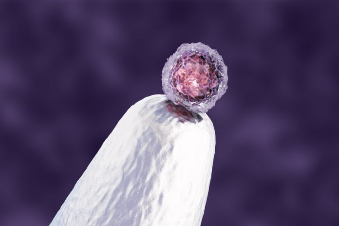
At the point of conception, the single-celled zygote (fertilised egg), through multiple subsequent rounds of cell division and the process of embryonic development, can form any specialised cell, and group into tissues found in the adult body. After about five days of cell division, the zygote forms a hollow ball of cells (about the size of a full-stop) known as a blastocyst; embryonic stem cells on the inside are ‘pluripotent’ and capable, through normal development, of giving rise to all the specialised cells found in the human body. During early embryo development, cells in different regions of the blastocyst form three cellular ‘germ layers’ (endoderm, mesoderm and ectoderm).
In each germ layer, these cellular descendants of pluripotent stem cells are already on their path to becoming a diverse array of highly specialised cells in different parts of the adult body. While all the genes in the human genome (about 22,000) are present in the nucleus of each cell, during this process of cellular ‘differentiation’ the protein products of certain regulatory genes (called transcription factors) can turn on, turn off, or otherwise alter the level of activity of numerous other genes (just like a single switch or dimmer can be used to control the output from multiple lights).
However, for most cells, once they are on their path to tissue specialisation, there is no turning back. As development progresses, cells become increasingly differentiated and specialised to the tissue environment in which they find themselves. Some adult stem cells (somatic) persist, but the cellular descendants of these part-differentiated are largely determined by their tissue environment. While the discovery of tissue-specific stem cells was an important advance, such cells are difficult to locate and hard to maintain in laboratory cultures.
Because of their enormous potential for use in regenerative medicine, such as alleviating some problems of ageing and in the treatment of chronic diseases, research and development (R&D) into stem cells is a global multibillion-dollar industry. According to one report, the field of regenerative medicine is expected to have a global market value of $53.7 billion by 2021. The 2018 governmental report from the Office for Life Sciences (the application of biology and technology to health improvement) showed that in the UK alone the sector had an approximately £70.3 billion in annual turnover, increasing greatly after factoring in supply chains. With one of the strongest and most productive health and life sciences industries in the world, the UK Government, through the Life Sciences Industrial Strategy paper committed nearly £500 million of investment.
For experimental research, human embryonic stem cells can be obtained from early-stage embryos, normally those formed in the laboratory but no longer required for assisted reproduction purposes, but this source comes with ethical and legislative issues; even words associated with the process (harvesting, cultured, laboratory, etc.) can be emotive and sound sinister to some. The requirement for a steady flow of new human embryos for research purposes has partly been addressed with an important development: the culturing of embryonic stem cells in the laboratory and their maintenance as cell-lines which retain their developmental flexibility.
Monthly advances are being made to better understand the cellular differentiation process using these laboratory cultures; the controlled differentiation of stem cells being an important requirement if they are to reach their full potential in regenerative medicine. Complementing this research, there are developments in genomics, particularly epigenomics (important for understanding gene regulation and expression) and transcriptomics (those genes actively being expressed in a particular cell or tissue, or at a particular time or set of circumstances).
In 2016, genomics-related activities in UK Life Science companies had an estimated £1 billion turnover and employed 1,800 workers. A further boost to stem cell R&D came in October 2016, with the launch of the Human Cell Atlas Project (HCAP), a global effort connecting biologists, clinicians, technologists, physicists, computational scientists, software engineers, and mathematicians.
HCAP aims to produce “a unique ID card for each cell type, a three-dimensional map of how cell types work together to form tissues, knowledge of how all body systems are connected, and insights into how changes in the map underlie health and disease”. A realistic goal considering the rapid advances in single-cell genomics; genomics, epigenomics, transcriptomics – all at the level of a single cell. In December 2018, the UK’s Medical Research Council announced that they would be investing £6.7 million to support the Human Cell Atlas initiative.
Dolly the sheep (often incorrectly thought of as the first cloned mammal) was born in the summer of 1996 following a process of nuclear transfer; the nucleus from a mammary gland cell of an adult sheep was transferred into an enucleated unfertilized egg cell from a different sheep. The ‘embryo’ developed in the laboratory and, once at a blastocyst stage it was implanted into a surrogate mother sheep and allowed to develop naturally to term. The real and lasting significance of Dolly was that it was the first mammal to be cloned from a fully differentiated adult somatic cell.
Since Dolly, there have been enormous advances in mammalian cloning via nuclear transfer, but for most species, the process is still very inefficient, and embryos produced via this cellular reprogramming process often show abnormal development. The real legacy of Dolly the sheep has not been primarily in advancing research into the cloning of mammals, but in highlighting the exciting prospect that differentiated cells may, in fact, be reprogrammable.
Researchers are making monthly advances in our understanding of how differentiated body cells can be reprogrammed to produce what are called ‘induced pluripotent stem cells’ (IPS) which subsequently, through controlled differentiation, can be grown back into any tissue. IPS are, therefore, differentiated somatic (body) cells that have been reprogrammed to behave like they were embryonic stem cells. IPS are an important research development, further reducing the demand for embryonic stem cells derived from human blastocysts and enabling scientists to better understand the normal development and cellular differentiation process. One thing’s for sure, these are all necessary and important steps along the road if we are to see stem cells becoming widely used in a cost-effective way for regenerative medicine and in the improvement of human quality of life with a steadily increasing average life expectancy.
The human body has a naturally low regenerative potential, but millions of people would benefit globally if tissues and organs could be replaced on demand. Traditionally, this has been done by organ transplantation, but demand greatly outstrips supply. Bringing together advances in stem cell research with those in the field of three-dimensional (3D) printing enhances the prospect of ‘3D bioprinting’. Artificial organ printing, by reprogramming patient-specific cells (IPS) and bioprinting them onto natural or biocompatible scaffolds, takes the prospect for regenerative medicine to another level. While many challenges remain, advances in stem cell R&D have been so rapid over the last decade that such ‘futuristic’ scenarios are perhaps not as distant as they might seem.
Graham Rowe
Lecturer in Biological Sciences
University of Derby
Tel: +44 (0)1332 590 500


