Open Access Government produces compelling and informative news, publications, eBooks, and academic research articles for the public and private sector looking at health, diseases & conditions, workplace, research & innovation, digital transformation, government policy, environment, agriculture, energy, transport and more.
Home Search
glaucoma - search results
If you're not happy with the results, please do another search
Peripapillary Intrachoroidal Cavitation, a masquerade of normal-tension glaucoma
Dr Adèle Ehongo discusses peripapillary intrachoroidal cavitation (PICC), a masquerade of normal-tension glaucoma.
Glaucoma clinic within the ophthalmology department
Professor Adèle Ehongo discusses her work in context of the ophthalmology department at Brussels University Hospital.
High-tech breakthrough could revolutionize glaucoma diagnosis and treatment
A groundbreaking new tool, The Glaucoma Field Defect Classifier (GFDC), developed by eye specialists at Addenbrooke’s Hospital, could transform glaucoma diagnosis and help change the way its managed worldwide.
New markers in blood could predict vision loss risk in glaucoma patients
Researchers from UCL and Moorfields Eye Hospital have identified potential markers in the blood that could change the treatment of glaucoma, a leading cause of irreversible blindness worldwide.
World Glaucoma Week: The importance of eye examinations
In recognition of World Glaucoma Week, The Association of British Dispensing Opticians (ABDO) highlights how a simple eye test is a key way to detect Glaucoma.
World Glaucoma Week 2020: A guide for contact lens wearers
As part of World Glaucoma Week, Alastair Lockwood, ophthalmologist and eye health specialist at Feel Good Contacts, explains how to minimise the risks of developing glaucoma in later life, and for lens wearers, how to ensure you are wearing them most effectively.
Peripapillary intrachoroidal cavitation and myopic peripapillary changes
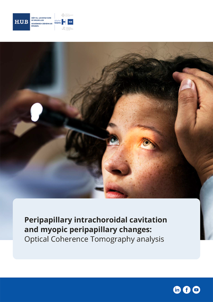
Peripapillary intrachoroidal cavitation and myopic peripapillary changes: Optical Coherence Tomography analysis.
Understanding the link between PICC and myopic complications
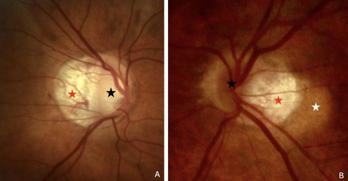
Dr Adèle Ehongo discusses the pathogenesis of peripapillary intra-choroidal cavitation and its implications for myopic complications.
Spotting peripapillary intra-choroidal cavitation using OCT
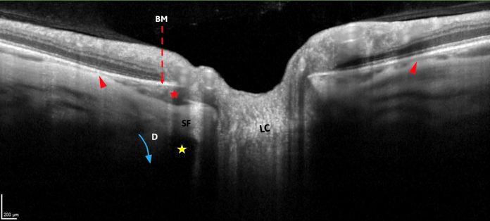
Adèle Ehongo explores the potential of optical coherence tomography for diagnosing peripapillary intra-choroidal cavitation in myopic eyes.
Adèle Ehongo MD, PhD – Hôpital Erasme
I am currently head of the glaucoma clinic at the Université Libre de Bruxelles (ULB), Hôpital universitaire de Bruxelles (HUB), CUB Hôpital Erasme, Bruxelles, Belgium.
I graduated as a doctor in 1992 and as ophthalmologist in 1997 from Université Libre de Bruxelles.
In my daily activity, my involvement in glaucoma, this...
The basics of myopia: What you need to know
Myopia has a significant economic and societal impact globally, and its prevalence in the digital age is increasing. We discuss the causes, symptoms and treatment for this condition.
Eye health: Understanding childhood myopia
Professor Nicola Logan, Professor of Optometry & Physiological Optics at Aston University, helps us to understand childhood myopia (short-sightedness), stating that early management in this vein is crucial for eye health.
Ultrananocrystalline diamond coating (UNCD™): Revolutionizing surface engineering
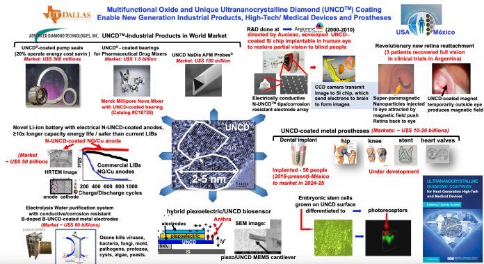
Unique, low-cost ultrananocrystalline diamond (UNCD™) coating is facilitating new generations of industrial products, high-tech devices, medical devices, and prostheses.
Unique Low-Cost Ultrananocrystalline Diamond Coating
Unique Low-Cost Ultrananocrystalline Diamond (UNCD™) Coating, enables New Generations of Industrial Products, High-Tech Devices, Medical Devices, and Prostheses.
What is age-related macular degeneration?
Age-related macular degeneration is known to affect millions of people around the globe and is fourth on the list of diseases that commonly lead to blindness, behind cataracts, preterm birth and glaucoma
Macular degeneration, also called age-related macular degeneration (AMD), is a medical condition that results in blurred or total...
Improving AI/ML services for ophthalmology and medicine
Eric Buckland, PhD of Translational Imaging Innovations, delves into how we can achieve better transparency, traceability, and reproducibility in AI/ML for ophthalmology and medicine.
Study finds biomarker for early multiple sclerosis diagnosis
Researchers have discovered that measuring retinal layer thickness can significantly improve the diagnosis of multiple sclerosis (MS).
1 in 4 people in the UK suffer from dry eye, but what is...
Although 17 million people are thought to be suffering from dry eye, the condition is not always easily diagnosed. How can we better understand it?
Ultrananocrystaline diamond (UNCD™) coatings for new generations high-tech/ medical devices/prostheses
Materials science, integration strategies, properties and more for the unique biocompatible Ultrananocrystalline Diamond (UNCD™) coating.
What are the causes and symptoms of diabetic retinopathy?
Diabetic retinopathy is the real medical term for diabetic eye disease. It is the most common cause of blindness in people of working age. 94 million people are affected worldwide.


