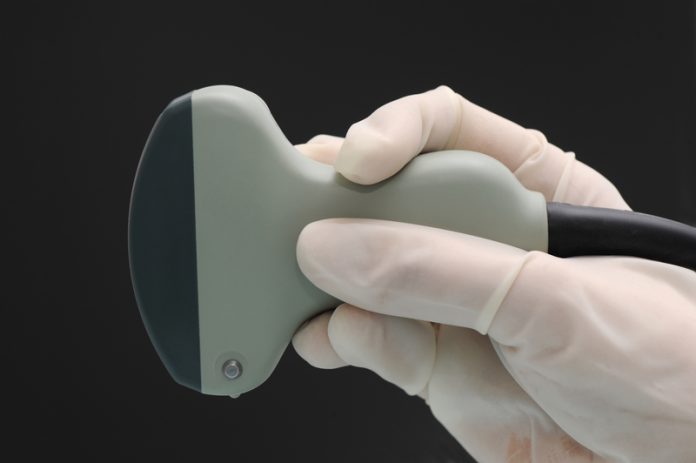
Virginia M Stewart at FESPA outlines the importance of guidelines for delivering emergency ultrasound treatment
As per the August 2012 American College of Emergency Physicians (ACEP) Emergency Ultrasound (EUS) section: “There are no definite protocols for training that have come from their distinct specialties (as of yet), therefore section members have employed the general ACEP training guidelines when applied to credentialing of mid-level providers. For nurses, many programs have successfully implemented training in IV placement and bladder volume assessment.”
In 2008, ACEP guidelines for non-resident emergency providers using ultrasound recommended:1
- A minimum of 4 hours of didactic training;
- A minimum of 150 ultrasounds reviewed by the designated reviewer;
- 25 ultrasounds in each of the 6 main areas of competency to obtain “general EUS competency”;
- A recommended 25 reviewed ultrasounds in any area of competency;
- 10 reviewed ultrasounds for procedures using ultrasound guidance.
Evidence for APP use of EUS applications
While many studies evaluate the performance of emergency physicians (EPs) trained in EUS, studies have only recently been emerging that specifically evaluate the EUS performance of advanced practice providers (APPs). In many academic programs, emergency APPs learn ultrasound alongside residents and EUS naïve attending physicians. In one prospective study, APPs post-test performance after a condensed EUS course was equivalent to the average score for all participants, and slightly exceeded scores of attending physicians.2 Areas of competency included: echocardiography, aorta, ectopic pregnancy, trauma, renal, gall bladder, and obstetric. Another prospective single-center study demonstrated an overall diagnostic accuracy or 85% for diagnosis of deep venous thrombosis with a 3 point scan protocol among 183 low-risk patients.3
Prompt recognition and treatment of tension pneumothorax is critical in a trauma setting and is within the scope of APPs. After a brief presentation, ultrasound naïve APPs accurately detected pneumothoraces, as well as normal hemothoraces, with a sensitivity of 95.4% (95% CI 0.75-0.99), a specificity of 100% (95% CI 0.81-1), a PPV of 100% and a NPV of 95.6%.4 Pre-hospital studies have demonstrated adequate EUS image quality and interpretation by non-physician providers in the setting of trauma, cardiac arrest, and thoracic ultrasound.5,6
Scope of practice of EUS for APPs
Point-of-care ultrasound is the use of ultrasound technology by trained providers for the bedside evaluation, diagnosis, treatment, and resuscitation of emergent, urgent, and ambulatory complaints. Typically EUS involves limited studies that answer a focused yes/no question pertaining to a specific organ system, and is performed by a physician, or APP under direct physician guidance.
EUS can be classified by the following functional categories:1
- Resuscitation;
- Diagnostic;
- Symptom or sign-based: ultrasound based on clinical pathways based on the patient clinical complaint, symptom or sign;
- Procedure guidance;
- Therapeutic and monitoring.
Applications of EUS1
- Trauma;
– Focused Abdominal Sonography in Trauma protocol (FAST).
- Intrauterine pregnancy;
– Identification of in all trimesters, and recognition of ectopic pregnancy.
- Abdominal Aortic Aneurysm (AAA) diagnosis;
– Diagnosis of aortic dissection.
- Cardiac;
– Limited: to include presence of cardiac motion during cardiac arrest, and identification of pericardial effusion and tamponade physiology;
– Advanced applications: qualitative and quantitative assessment of ejection fraction, focal wall motion abnormalities, assessment of valvular abnormalities and pathologies, identification of thrombus and cardiac masses, and identification of lethal and non-lethal arrhythmias including electromechanical dissociation;
– Procedural guidance for pericardiocentesis.
– Evaluation of traumatic, infectious, and neoplastic pulmonary processes, thoracentesis and thoracostomy procedural guidance, and mediastinum evaluation.
- HEENT;
– Confirmation of endotracheal tube placement, diagnostic angioedema classification, evaluation of vocal cord injury, peritonsilar abscess identification and procedural guidance of drainage, limited thyroid evaluation.
- Biliary;
– Diagnosis of cholecystitis, cholelithiasis, and pancreatitis/choledocolithiasis.
- Renal/urinary tract;
– Bladder volume quantification, identification of hydronephrosis.
- Deep venous thrombosis diagnosis (limited compression study);
- Soft tissue and musculoskeletal;
– Diagnosing cellulitis, necrotizing fasciitis, and other soft tissue infection, procedural guidance for abscess drainage, and foreign body removal, diagnosis of long bone fracture, diagnosis and procedural guidance for joint and fracture reduction and arthrocentesis.
- Ocular;
– Diagnosis of retinal detachment, vitreous hemorrhage, foreign body, lens dislocation, globe rupture, and central retinal artery and venous occlusion.
- Procedure guidance;
– Vascular access, peripheral nerve blocks, and any invasive procedure performed in the emergency department.
- Bowel;
– Including diagnosis of intussusception, appendicitis, pyloric stenosis, diverticular disease, volvulus, obstruction.
- Adenexal pathology diagnosis;
– Infectious more so than neoplastic.
- Testicular;
– Diagnosis and procedural guidance for torsion.
Level of Supervision
Many academic and community health systems within the United States allow for the independent performance and interpretation of EUS by APPs, with direct visualization, or immediate over-read and verification by the attending physician. Depending on the level of training, facility, and patient acuity, the level of supervision (direct or indirect) varies. Direct supervision involves the attending EP in the room during the ultrasound. Indirect supervision involves the EP immediately reviewing images obtained. It is this author’s recommendations that direct attending physician supervision be mandatory for all unstable patients, and procedural guidance involving major vascular structures or risk of significant complication to major vascular or other structures.
EUS in stable patients with low pretest probability of significant morbidity or mortality, and “low risk” procedures (superficial abscess incision and drainage, foreign body identification or removal, and nerve blocks that do not involve risk to significant vascular structures) may be supervised indirectly, with the attending EP present for key parts of the procedure, and reviewing images obtained immediately after they are taken and prior to the patient disposition.
1 Emergency Ultrasound Guidelines, 2008 ACEP policy statement
2 Mandavia, D. P., Aragona, J., Chan, L., Chan, D., & Henderson, S. O. (2000). Ultrasound training for emergency physicians – A prospective study. Academic Emergency Medicine : Official Journal of the Society for Academic Emergency Medicine, 7(9), 1008-1014. doi:10.1111/j. 1553-2712.2000.tb02092.x
3 Kline, J. A., O’Malley, P. M., Tayal, V. S., Snead, G. R., & Mitchell, A. M. (2008). Emergency clinician-performed compression ultrasonography for deep venous thrombosis of the lower extremity. Annals of Emergency Medicine, 52(4), 437-45. doi:10.1016/j.annemergmed. 2008.05.023
4 Monti, J. D., Younggren, B., & Blankenship, R. (2009). Ultrasound detection of pneumothorax with minimally trained sonographers: A preliminary study. Journal of Special Operations Medicine : A Peer Reviewed Journal for SOF Medical Professionals, 9(1), 43-6.
5 Roline, C. E., Heegaard, W. G., Moore, J. C., Joing, S. A., Hildebrandt, D. A., Biros, M. H., . . . Reardon, R. F. (2013). Feasibility of bedside thoracic ultrasound in the helicopter emergency medical services setting. Air Medical Journal, 32(3), 153-7. doi:10.1016/j.amj.2012.10.013
6 Byars D, Stewart V, Knapp B. Emergency Medical Services Focused Assessment with Sonography in Trauma and Cardiac Ultrasound in Cardiac Arrest: The Training Phase. Oral presentation of abstract. Society of Academic Emergency Medicine. May 2012.
Virginia M Stewart
Forsyth Emergency Services, PA
P.O. Box 25447
Winston-Salem
NC 27114
Please note: this is a commercial profile


