Open Access Government produces compelling and informative news, publications, eBooks, and academic research articles for the public and private sector looking at health, diseases & conditions, workplace, research & innovation, digital transformation, government policy, environment, agriculture, energy, transport and more.
Home 2026
Archives
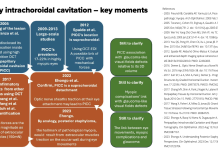
Peripapillary intrachoroidal cavitation and biomechanical considerations: A multi-stage narrative
Dr Adèle Ehongo discusses Peripapillary intrachoroidal cavitation and biomechanical considerations in a multi-stage narrative.
Ocular nutrition for a digital generation
Effective nutritional solutions to support healthy vision in children affected by Computer Vision Syndrome (CVS). Discover how healthcare professionals and policymakers can promote eye health and reduce the impact of screen time on kids.
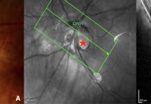
Peripapillary Intrachoroidal Cavitation, a masquerade of normal-tension glaucoma
Dr Adèle Ehongo discusses peripapillary intrachoroidal cavitation (PICC), a masquerade of normal-tension glaucoma.
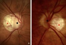
Understanding the link between PICC and myopic complications
Dr Adèle Ehongo discusses the pathogenesis of peripapillary intra-choroidal cavitation and its implications for myopic complications.
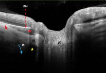
Spotting peripapillary intra-choroidal cavitation using OCT
Adèle Ehongo explores the potential of optical coherence tomography for diagnosing peripapillary intra-choroidal cavitation in myopic eyes.
Sight loss research: Looking forward to an equitable future
Keith Valentine, Chief Executive of Fight for Sight and Vision Foundation, shares how these newly merged organisations are driving efforts in sight loss research to improve patient care.
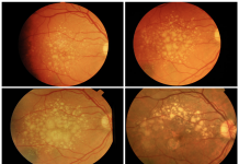
Understanding age-related macular degeneration
Tunde Peto, Professor of Clinical Ophthalmology at Queen’s University Belfast, describes the symptoms, causes and treatments for age-related macular degeneration and how the prevalence of the disease could be reduced.

