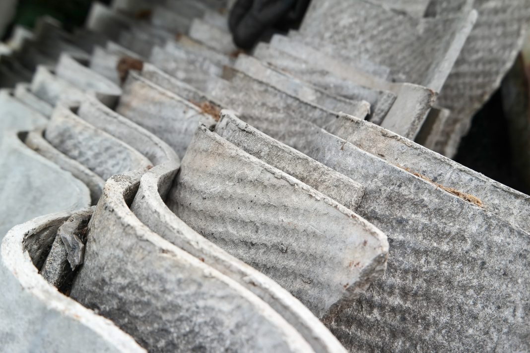
Jean Pfau and Kinta Serve explore a critical and novel hypothesis concerning the size of fibers in asbestos disease pathogenesis
The strange definition of asbestos
A mineral fiber is defined as having an aspect ratio (length/width) that is greater than three, but the mineral is only considered asbestos if it also has certain commercially-valuable qualities like durability and tensile strength. Only six silicate-based commercial minerals are officially called ‘asbestos’. However, currently, to be counted as asbestos, a fiber also must be long enough to be seen with light microscopy. Why?
A perspective on smallness
In order to remain airborne as dust and inhaled into the lungs, where they can cause disease, asbestos fibers must be extremely small, in the range of micrometers (μm = 0.001mm). Historical studies evaluating optimal ways to detect and count asbestos fibers used the best technology available at the time: phase contrast light microscopy (PCM). The counting process is challenging, so rules were established to standardize the process for the entire industry. Due to the resolution limits of the instruments, to be counted, a fiber had to be >5μm long.
A great deal of work has now shown that the vast majority of fibers in commercial exposures are very tiny (<5μm long and <1μm wide). (1) Also, wind-blown environmental dust from asbestos-containing soils and rock contains primarily these tiny fibers, which are extremely difficult, often impossible, to see with PCM and therefore require the tedious, expensive process of electron microscopy (EM). So, in a commercial setting, the 5μm length limit was generally adequate for determining the presence and type of asbestos, but it had nothing to do with health effects.
It has long been erroneously believed that tiny fibers (<5μm long) are cleared from the lung by the ‘mucociliary escalator’, a constant movement of mucus up the larger airways by cilia. While it is true that this process effectively clears some fibers, long and short, tissue fiber burden studies using EM have revealed millions of very short, thin fibers in all thoracic and abdominal tissues of patients who had died from asbestos-related diseases. (5,6) By current counting rules, these fibers would not be counted as asbestos, but are they really harmless?
Counting rules ignore short fibers partly because misinterpretation of studies by M.F. Stanton and others led people to believe that only long (>8μm) fibers cause disease. (2) However, careful reading of those studies questions that belief. (1,2,3) Asbestos causes several diseases, but little is known about what characteristics of asbestos, such as length or mineralogy, cause a particular disease. Mesothelioma risk may increase with higher doses of long fibers. (4) However, less is known about other, more common, asbestos-related diseases.
Systemic autoimmune disease
One of the lessons from Libby, Montana, was that, in addition to lung diseases, amphibole asbestos causes systemic autoimmune diseases. (7,8,9) Libby Amphibole asbestos co-existed geologically with vermiculite mined for decades near Libby, Montana, and therefore remained a toxic contaminant of the ore and commercial products. To cause autoimmune disease, two things must happen, and both require tiny fibers. First, the fibers must leave the lung via the lymphatics, a system of tiny vessels that carry lymph to and from tissues and blood. Studies have demonstrated that this does happen and that the average length of fibers in the pleura and lymph nodes is 2.5μm, and most are too narrow (<0.2μm) to be detected by light microscopy. (3) EM is required to find these fibers, which end up in lymph nodes and virtually all tissues throughout the body. (5) Second, the fibers are engulfed by several cell types, most often the macrophage. The macrophage’s job is to engulf foreign material, enter the lymphatics, and alert the immune system to their presence. Macrophages are about 10μm in diameter, making it impossible to engulf fibers longer than 5-8μm. Macrophages containing engulfed particles have been found in tissues outside the lung and are associated with lymphatic tissue. (3) This shows that systemic immune activation does occur and that tiny fibers are responsible.
Experimental comparisons of short and long asbestos
Early evidence supporting the belief that only long fibers are dangerous came from studies that counted fibers (using light microscopy) present in lung lesions and found primarily long fibers. However, EM studies revealed that for every long fiber, many tiny fibers are also present. (1,3) Several innovative studies used amosite asbestos samples that were either left alone or ground into finer particles. (10) While the idea was clever, the approach was flawed. The unground amosite (called Long Fiber Amosite (LFA)) was shown to be highly carcinogenic, while the ground sample was minimally so, leading the researchers to conclude that only long fibers are pathogenic. However, LFA contained a high percentage (70%) of fibers < 5μm in length. (1,11) So, it was NOT a sample of long fibers: it was a mixture of long and short. In addition, the grinding process produced primarily non-fibrous particles and changed the surface characteristics enough that these studies actually compared those variables, not fiber length. (11) Nevertheless, elegant studies have teased apart details of the responses to LFA and SFA, showing that while both affect cells and tissues, they activate distinct pathways, (12) suggesting that they could work together to produce disease.
Reviews of the literature reveal that studies using samples enriched in short fibers consistently demonstrate health effects. (1, 2) Long fibers seem to initiate inflammation and carcinogenesis, but short fibers involve the immune system, thereby leading to immune dysregulation and non-resolving inflammatory pathways. The immune system then can no longer switch over to healing mode to stop fibrosis, control cancer, or avoid autoimmune reactions.
Therefore, a novel hypothesis is that asbestos disease pathogenesis requires a combination of both long and short fibers. While clearly more research is needed, perhaps all fibers should be counted in exposure testing. Thank you, Libby, Montana, for your critical lessons on asbestos! (9)
Funded by ATSDR screening grant 5 NU61TS000295-05-00
References
- Boulanger 2014, http://www.ehjournal.net/content/13/1/59
- Militello 2021, https://doi.org/10.3390/min11050525
- Dodson 2003, https://onlinelibrary.wiley.com/doi/abs/10.1002/ajim.10263
- Wylie, 2023, https://doi.org/10.1016/j.envres.2022.114688
- Oberdorster 1988, https://doi.org/10.1093/annhyg/32.inhaled_particles_VI.149
- Caraballo-Arias 2023, https://doi.org/10.23749/mdl.v114i6.14946
- Pfau 2005. https://doi.org/10.1289/ehp.7431
- Diegel 2018. https://doi.org/10.1080/15287394.2018.1485124
- Pfau 2024, https://www.openaccessgovernment.org/article/lessons-from-libby-understanding-the-impact-of-asbestos-exposure/171870/
- Davis 1986, https://pubmed.ncbi.nlm.nih.gov/2872911/
- Riganti 2003, https://doi.org/10.1016/S0041-008X(03)00339-9
- Leinardi 2023, https://doi.org/10.3390/ijms242015145

This work is licensed under Creative Commons Attribution-NonCommercial-NoDerivatives 4.0 International.


