Ramani Ramchandran, Professor at the Medical College of Wisconsin, investigates ciliary biomarkers for diagnosing and prognosing vascular health
Vessels carry blood and biomarkers, which are substances in blood that inform the underlying status of vascular health in people. To date, most biomarkers in the blood are associated with proteins, lipids, sugars, and nucleic acids. Cellular organelles are rarely considered as biomarkers. Our work on a tiny hair-like organelle called cilia found in most cells and the proteins inside them as potential biomarkers that inform underlying vascular health (1) is one of the first studies to consider this concept.
Cilia as blood flow sensors
Cilium (singular, plural cilia) comes in two varieties: non-motile primary cilium and motile cilia. Each cell in the body is thought to have one primary non-motile cilium that is continuously formed and reformed during cell’s life cycle. Occasionally, some cells in the body have more than one cilium. However, the purpose of having multiple cilia is not fully clear. Blood vessels are like hollow tubes that carry blood inside them. Cilia are found in both arteries and veins that carry blood, and in lymphatic vessels that carry lymph fluid.
Endothelial cells that form the underlying surface of the blood vessel contact (2) blood flowing inside the tube. Cilium were originally thought as blood flow sensors, and by sensing blood flow, calcium molecules get released along with nitric oxide, a gas that promotes dilation of blood vessels. Thus, cilia can convert mechanical forces into chemical reactions inside a cell, a process called mechanotransduction.
Cilia length and flow
Blood vessels with low fluid force have longer endothelial cilia, while blood vessels with high fluid force have shorter endothelial cilia or no cilia. Thus, in regions of low flow, cilia are enriched. For example, in the aortic arch vessel of the heart, cilia are in the curved portion of the vessel where the flow is low, and interestingly is the region prone for plaque buildup (atherosclerosis). Researchers have indeed identified that endothelial primary cilia prevent atherosclerosis. (2)

Cilia assembly and disassembly
In 2001, Iomini et al., in a paper in the Journal of Cell biology (3), first reported that primary cilia of human umbilical vein endothelial cells disassembled under high flow conditions. In 2019, Tim Stearns group at Stanford showed that primary cilia loss in mammalian cells occurred by the whole cilia shedding. (4) Others have shown that partial ciliary fragments are observed in urine during acute kidney injury. (5) These studies gave birth to the notion that cilia assembly and disassembly are important processes that occur during flow changes and organ injury. Importantly, whole cilia fragments or parts of them were observed in body fluids.
Cilia deciliation in the vasculature
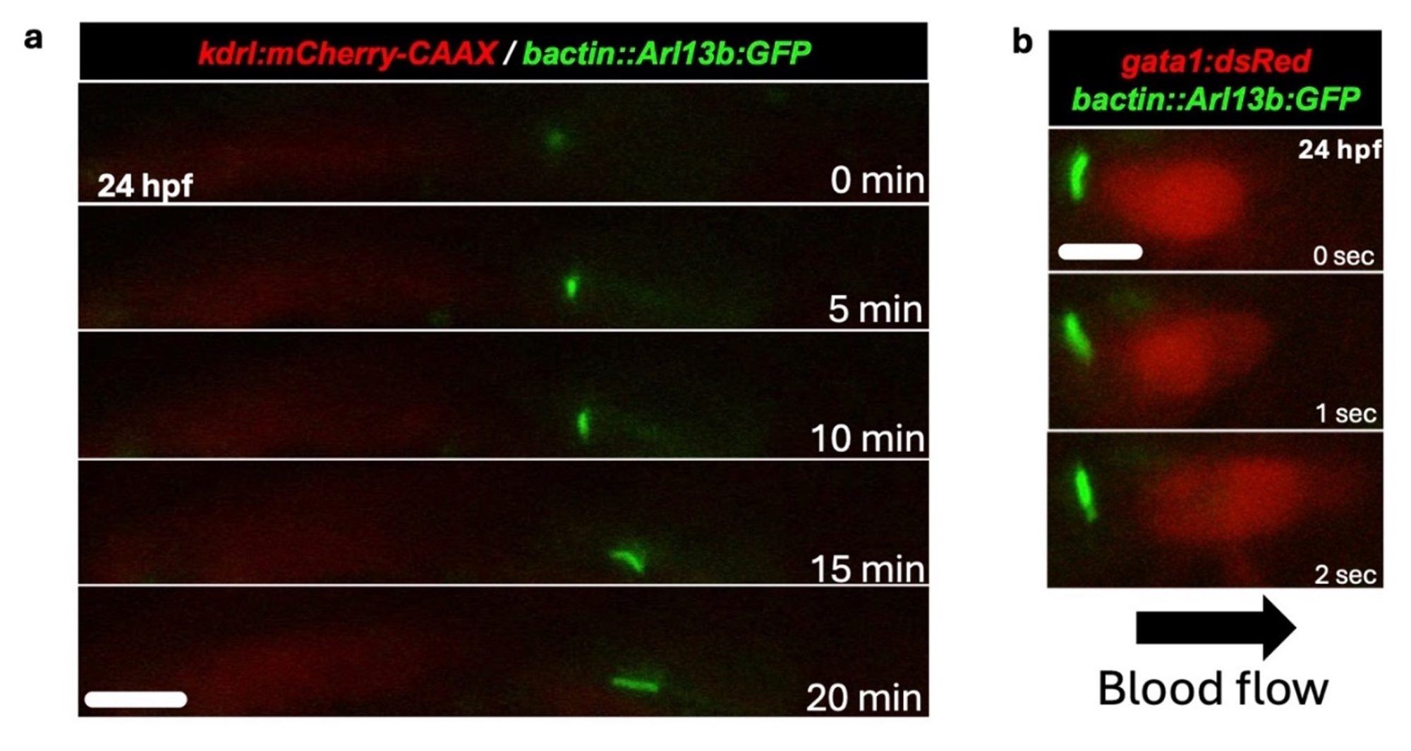
Based on the observation that cilia disassemble upon high flow, our group hypothesized that if cilia are disassembled, then ciliary fragments and the proteins inside them should also be released and be detectable in body fluids, including blood. We tested this hypothesis in the 2022 JCI Insight paper from our group where, using primary endothelial cell culture, we showed that, indeed, supernatants from primary endothelial cells grown in culture and subjected to high flow conditions displayed ciliary fragments and proteins. (1)
We also demonstrated in vivo in the freshwater zebrafish model system that when they were subjected to high flow conditions, cilia disappeared from their vessels, and ciliary protein levels were lower on zebrafish endothelial cells.
Cilia detection in human samples
We then asked, what about human samples? Can we detect ciliary proteins in human samples? To answer this question, we focused on how flow might affect the removal of cilia from endothelial cells. We hypothesized that red blood cells in human blood will interact with cilium on endothelial cell surface to dismantle them.
To test this hypothesis, we procured human blood samples from sickle cell patients who had deformed red blood cells (sickle in shape) that affected the blood flow and investigated them for cilia proteins. To our amazement, sickle red blood cells have high amounts of ciliary protein and procure more cilia upon interaction with endothelial cells in culture under flow conditions. The underlying signals that mediate the red blood cell-triggered removal of endothelial cell cilia is partly oxidative stress.
Ciliary biomarkers as tools for detecting the underlying vascular state
What does all this mean, and where do we go from here? For now, we have been able to detect ciliary proteins in serum, plasma, urine, and cerebrospinal fluid. This means the opportunity to develop them as biomarkers of underlying vascular health condition is prime. We have indeed embarked on this journey and formed a start-up company (CIAN, Inc.) that is developing ciliary biomarkers for brain injury and other conditions. The main roadblocks that need overcoming are assessing the clinical value of the ciliary protein changes and the extent of clinical intervention that will materialize from this information.
Also, as cilia are expressed in most vascular beds (cerebral, pulmonary, coronary), the ability to distinguish across these beds using flow-based biomarkers remain an open question. On the other hand, any condition where flow is compromised offers an opportunity for ciliary biomarker application, which enhances its value as a biomarker tool. At the very least, we posit that detecting ciliary proteins in blood or body fluids will serve as surrogates for evaluating disease states and thus offer the physician an additional tool in their quest to deliver optimal healthcare.
References
- Gupta A, Thirugnanam K, Thamilarasan M, Mohieldin AM, Zedan HT, Prabhudesai S, Griffin MR, Spearman AD, Pan A, Palecek SP, et al. Cilia proteins are biomarkers of altered flow in the vasculature. JCI Insight. 2022. doi: 10.1172/jci.insight.151813
- Dinsmore C, Reiter JF. Endothelial primary cilia inhibit atherosclerosis. EMBO Rep. 2016;17:156-166. doi: 10.15252/embr.201541019
- Iomini C, Tejada K, Mo W, Vaananen H, Piperno G. Primary cilia of human endothelial cells disassemble under laminar shear stress. J Cell Biol. 2004;164:811- 817. doi: 10.1083/jcb.200312133
- Mirvis M, Siemers KA, Nelson WJ, Stearns TP. Primary cilium loss in mammalian cells occurs predominantly by whole-cilium shedding. PLoS Biol. 2019;17:e3000381. doi: 10.1371/journal. pbio.3000381
- Kong MJ, Bak SH, Han KH, Kim JI, Park JW, Park KM. Fragmentation of kidney epithelial cell primary cilia occurs by cisplatin and these cilia fragments are excreted into the urine. Redox Biol. 2019;20:38-45. doi: 10.1016/j.redox.2018.09.017


