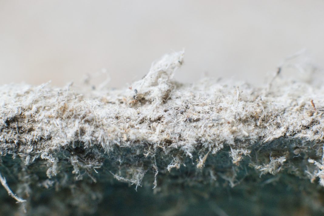
Karen Lee Morrissette and Jean C. Pfau from the Center for Asbestos-Related Disease discuss the clinical presentation and complexity of the autoimmune disease progression among those exposed to Libby Amphibole
The Center for Asbestos Related Disease (CARD) opened in 2000 to provide asbestos health screening and ongoing care for those exposed to Libby Amphibole asbestos at the Superfund site in Libby, Montana. A vermiculite mine, open for a seventy-year period ending in 1990 and at one time producing up to 80% of the world’s vermiculite supply, was naturally contaminated by asbestiform fibers – tremolite, winchite, and richterite – a combination now known as Libby Amphibole. Not only were mine workers exposed, but also their families and most of the nearby town of Libby. In 2009, the Environmental Protection Agency (EPA) declared the area an environmental public health emergency, the first designation of its kind in the US.
Of the three fibers found in Libby Amphibole, only the least prevalent, tremolite, is actually regulated by the government. Despite ongoing debate about whether non-regulated asbestiform fibers can cause disease, CARD’s twenty-year experience has proven that not only can they produce the classic malignant diseases such as mesothelioma and lung cancer but also a progressive non-malignant lung disease with pleural changes over and beyond those typically found with commercial chrysotile fibers, as well as interstitial disease. During the past decade, CARD and its research collaborators have attempted to tease out the science behind why Libby Amphibole exposure may result in a clinical presentation that differs from that classically taught. The answer, it seems, lies within the immune system.
Over time, CARD providers, as well as local pulmonologists and rheumatologists, became aware of an increased prevalence of autoimmune disease in those exposed to Libby Amphibole compared to the general population. In a 2018 article, Kalispell rheumatologist Roger Diegel and his colleagues found that 13.8% of the screened CARD population – then roughly 6600 – had been diagnosed with a systemic autoimmune disease compared to 10% in the general population. (1) While the estimates vary, women are roughly four times more likely to be diagnosed with an autoimmune disease than men. For Libby patients, the distribution was approximately 1:1. (1)
Systemic autoimmune disorders
Many Libby patients have been diagnosed with two, and sometimes three, different systemic autoimmune disorders. This likely results from the mixed symptom profiles common in the population. Diegel et al. found that elements of systemic lupus erythematosus (SLE), scleroderma, rheumatoid arthritis (RA), and undifferentiated connective tissue disorder were frequently seen. (1) Diagnoses of polymyalgia rheumatica, Sjogren’s Syndrome, and psoriasis with psoriatic arthritis have also become routine. This immune stimulation is not routinely seen in patients exposed to commercial chrysotile asbestos. The unusual symptom groupings can be confusing to providers looking for classical disease definitions, and as a result, these individuals often end up with either no diagnosis or multiple diagnoses.
There are subpopulations of those with asbestos-related lung disease due to Libby Amphibole who, after diagnosis, have a relatively benign course, while others, often with similar exposure, progress rapidly to a severely restrictive form of pleural fibrosis, with or without interstitial fibrosis. This latter group is at risk of death from respiratory failure. Current research suggests that this difference may be related to how each individual’s immune system reacts to Libby Amphibole. Asbestiform fibers enter the body, most commonly through inhalation, and are identified as foreign by the immune system. Unfortunately, the body is not able to break the fibers down. In those whose immune systems remain stimulated over time, constantly attacking these foreign particles, the persistent recruitment of macrophages and bombardment with immune system mediators and autoantibodies leads to a chronic state of inflammation. This ongoing inflammatory response, in turn, leads to collagen deposition, progressive scarring, and DNA mutations that can eventually lead to cancer. Inexplicably, others exposed to Libby Amphibole seem able to control this vicious cycle involving the immune system, tending to show minimal or at least a comparably slower disease progression.
Immune markers like antinuclear antibodies
Multiple studies led by Immunotoxicologists Dr Jean Pfau of Montana State University and Dr Kinta Serve of Idaho State University have shown an association between immune markers like antinuclear antibodies (ANA), anti-mesothelial cell antibodies (MCAA), and anti-plasminogen (anti-PLG) with progressive fibrosis. (2,3) As a result, CARD added ANA to its screening process in 2019 and anti-PLG in 2021. Using these markers benefits screeners in two ways. A positive ANA can be correlated with symptoms that may suggest unrecognized autoimmune disease, allowing for rheumatologic referral and work-up. Positive ANA or anti-PLG may also identify patients that, if diagnosed, should be monitored more closely for rapid disease progression.
The following case study elucidates the complexity of the disease presentation. A 50-year-old female, a non-smoking Libby native, presents for screening. She gives a history of joint pain and swelling diagnosed initially as osteoarthritis and later as seronegative rheumatoid arthritis. Because of some dry eye issues, another provider gave her a diagnosis of Sjogren’s Syndrome. She has spent the last thirty years working in a local plywood plant. Her childhood home had vermiculite insulation, and she often played in piles of vermiculite around town. She complains of increasing dyspnea over the last 5-6 years, in addition to cough and right-sided chest pain with exertion, making it difficult for her to continue working.
On pulmonary function testing (PFT), she has a moderately restrictive pattern, and on CT of the chest, there is moderate, thin, diffuse pleural thickening in the mid and lower chest, more so on the right. ANA is positive. She is diagnosed with asbestos-related lung disease. Over the next decade, she becomes unable to work due to increasing dyspnea and chest pain.
Her pleural fibrosis becomes thicker, eventually encasing the lungs, and she begins to develop some interstitial fibrosis. Ten years after diagnosis, she is oxygen-dependent, and her tolerance for activity is very low, resulting in significant weight gain and additional impairment of her breathing.
Future research
Future research could compare asbestos-related lung disease progression in patients on treatment for autoimmune disease versus those who are not. This could lead to clinical investigation into whether similar medications affecting the immune system might be useful in treating progressive pleural disease.
References
- Diegel, R., et al., 2018. https://www.tandfonline.com/doi/full/10.1080/15287394.2018.1485124
- Pfau JC., et al., 2019. https://www.tandfonline.com/doi/full/10.1080/08958378.2019.1699616
- Serve KM., 2013. https://www.tandfonline.com/doi/full/10.3109/08958378.2013.848249


