Sabine Mai and Aline Rangel-Pozzo, at the CancerCare Manitoba Research Institute and The University of Manitoba, Winnipeg, Canada, discuss genomic instability in relation to 3D spatial organisation of telomeres
Genomic instability and cancer
Genomic instability is a hallmark of cancer. It drives the evolution of cancer cells having a genome that differs from that of normal cells. It enables the development of cell clones and cell-to-cell heterogeneity within the tumour and between the tumour and its metastatic site(s).
The presence of genomic instability in cancer was first reported by David von Hanseman (1858-1920). His work as a pathologist in Virchow’s group in Berlin concluded that cells exhibiting genomic instability were not found in normal tissue but only in tumour tissues. His illustrations showed chromosomal instability, which included aberrant chromosome numbers (aneuploidy: gain or loss of chromosomes); and chromosomal missegregation between daughter cells.
The first mechanistic studies into genomic instability were performed by Theodor Boveri (1862-1915), whose work proposed that genomic instability led to the development of cancer and was not compatible with normal cell life. While he did not use the term ‘genomic instability’, his data clearly implicated it in this malignant process.
Genomic instability is a dynamic process and continues to evolve with every cell division. Therefore, if samples are taken at an early time point of tumour development and at later stages, genetic changes will have occurred.
The three-dimensional (3D) space of the nucleus: spatial genome disorder in cancer
The nucleus harbours genetic information. This information is not simply organised in a random manner but has a clear organisation. In normal cells, chromosomes assume specific places within the 3D space of the nucleus. The spaces occupied by the chromosomes are called ‘chromosome territories’. They have been conserved during evolution, and they are cell-type and differentiation-specific. This organisation has functional importance; for example, animals with night vision have a chromosome organisation in their rod cells that is distinct from the one found in animals with diurnal vision. Similarly, hepatocytes have a different arrangement of chromosome territories and chromosome neighbourhoods than lymphocytes.

The organisation of chromosomes and of the genes they harbour is key to transcriptional regulation. Genes located near the periphery of the 3D nucleus are commonly repressed, while genes found in the centre of the nucleus are expressed.
Cancer cells have re-ordered the genetic information within the nuclear space and often change the position and orientation of chromosomes, which enables new transcription patterns. Cancer cells also change the way their DNA is packaged and increase the presence of interchromatin space. Moreover, when using telomeres, the ends of chromosomes, as structural markers for genome (in)stability, cancer cells, compared to normal cells, show very significant changes in their 3D organisation.
3D telomere profiling in cancer
3D imaging of telomeres developed and carried out at the Genomic Centre for Cancer Research and Diagnosis (GCCRD) at the University of Manitoba and CancerCare Manitoba (Winnipeg, Canada) enabled the fine-mapping of nuclear organisation of the cancer cell genome. At nanoscale resolution, Dr. Mai’s team and collaborators visualised and measured the structural organisation of the cancer genome; telomeres were used as surrogate markers of genome organisation and proved to play a role as structural biomarkers of genomic instability.
Multiple cancers were examined. 3D telomere profiling allowed the team to quantify the level of genomic instability, the risk to progression and/or the response to treatment. Examples include lymphoid (such as, Hodgkin’s Lymphoma, Multiple myeloma, Chronic Myeloid Leukemia, Myelodysplastic Syndromes and Acute Myeloid Leukemia) and solid tumors (such as neuroblastoma, glioblastoma, thyroid cancer, and prostate cancer). The software developed by the team (TeloView® – currently proprietary to Telo Genomics Corp. Toronto, Canada) allowed for single-cell profiling and informed about each patient’s cancer.
As a result of these initial clinical studies, a biotech company, Telo Genomics Corp., was founded. Telo Genomics Corp. is located at the MaRS Discovery District in Toronto, ON, Canada (telodx.com). The company is dedicated to personalised medicine solutions in cancer, with a current focus on multiple myeloma.
An example of nuclear remodeling: nuclear remodelling and risk assessment in prostate cancer
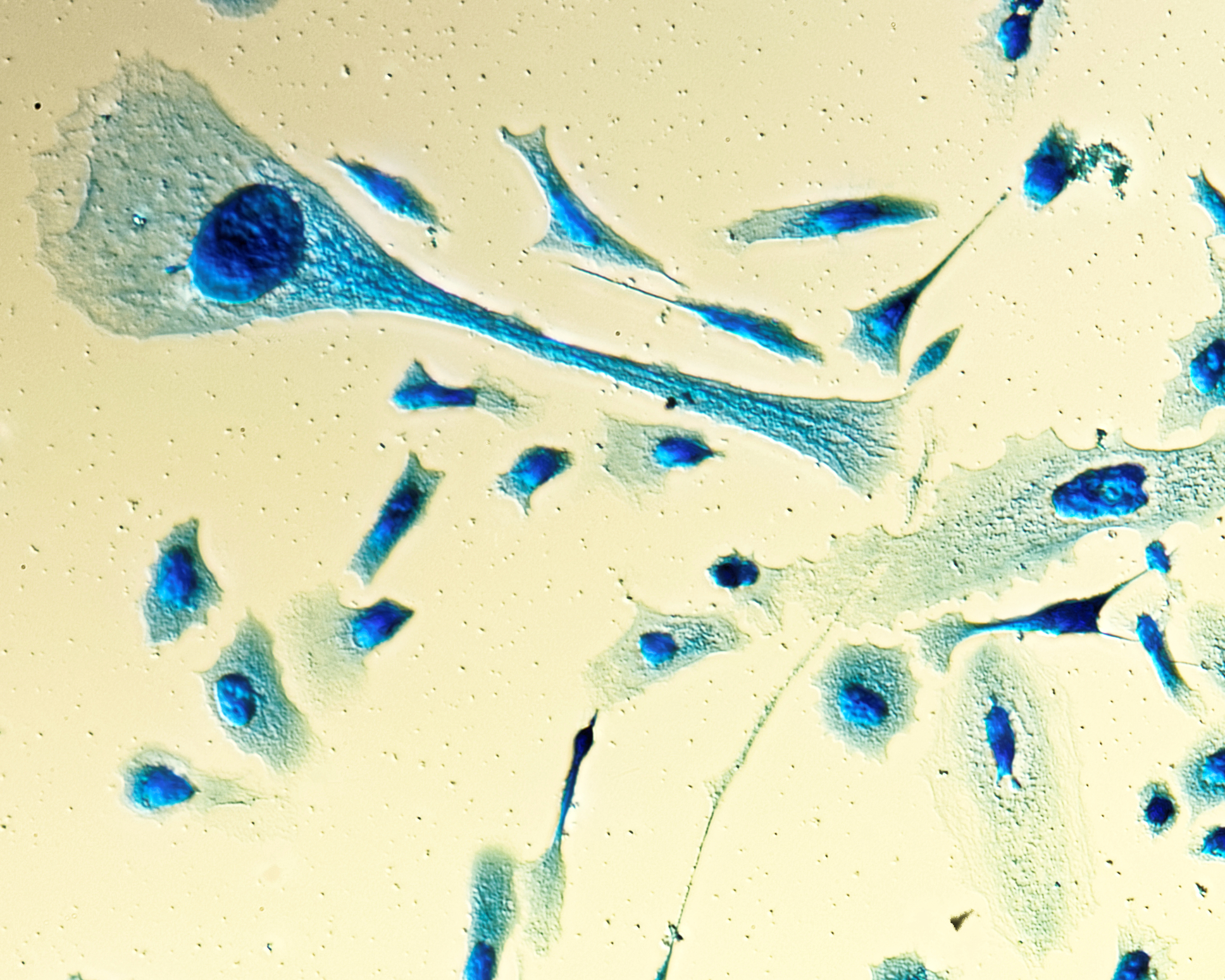
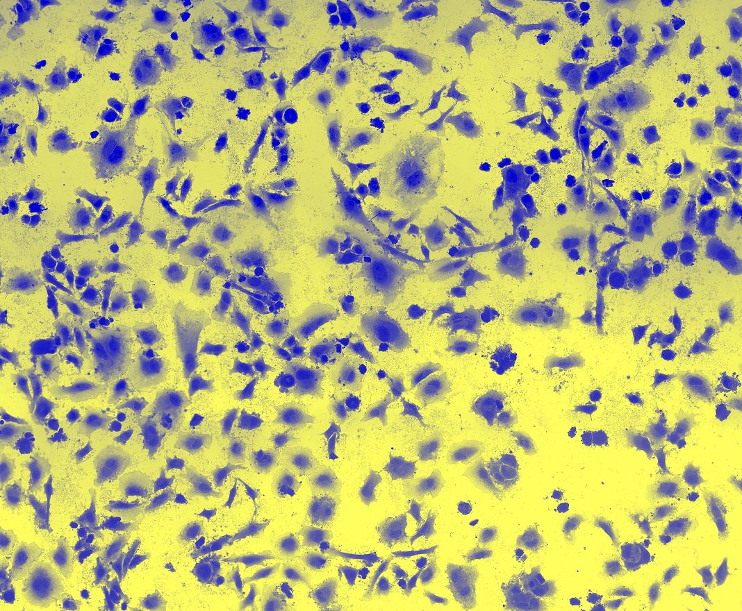
Prostate cancer is the 2nd most common cancer in men globally, with 1.4 million new cases in 2020. Intermediate-risk prostate cancer in men is often stable but sometimes aggressive. No clear test predicts which patient is with either type of prostate cancer. Research done by the Mai team, in collaboration with the urologists Drs. Drachenberg and Saranchuk at CancerCare Manitoba (Winnipeg, Canada) has focused on circulating tumor cells (CTCs) isolated from the blood of prostate cancer patients. Such isolates are called ‘liquid biopsies’, and these blood samples contain (CTCs) that originate from the tumor. Our analyses of 3D telomere profiling indicate that the genomic profile of CTCs in intermediate-risk prostate cancer predicts stability or aggressiveness of disease better than the commonly used prostate-specific antigen (PSA) or the Gleason score (Drachenberg et al., Cancers, 2019, Jun 20;11(6):855. doi: 10.3390/cancers11060855.PMID: 31226731). This study was funded by the Prostate Cancer Fight Foundation/Manitoba Ride for Dad.
The assessment of nuclear architecture
The assessment of nuclear architecture enables the analysis of genomic instability in cancer. Structural biomarkers, such as the spatial arrangement of cancer genomes, are poised to become future tools in personal patient risk assessment.

This work is licensed under Creative Commons Attribution-NonCommercial-NoDerivatives 4.0 International.


