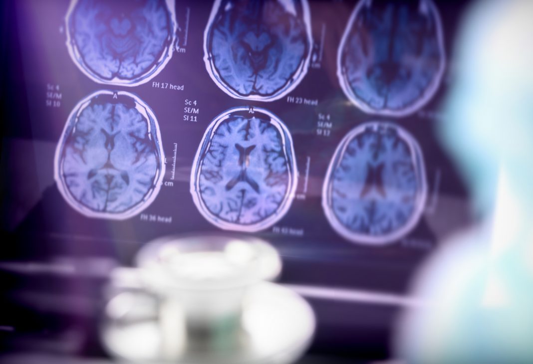
Effective treatments for amyloid-associated neurological diseases are desperately needed; H. Robert Guy, CEO of Amyloid Research Consultants, talks us through the obstacles and opportunities associated with structure-based drug design
The FDA recently approved an antibody drug, Lecanemab, aka Leqembi, that slows neuronal degeneration caused by Alzheimer’s Disease (AD). Granted, Lecanemab is no miracle drug: it is not a cure, it can have deleterious side effects, and it is expensive. Nonetheless, it is the first antibody treatment for AD to be efficacious. Lecanemab antibodies were developed to target amyloid beta (Aβ), long suspected of being a major cause of AD and a prime constituent of amyloid plaques. This development was a surprise since previous attempts to use antibodies to Aβ fibrils have failed.
Numerous researchers have long argued that Aβ fibrils are not the primary culprit; smaller Aβ assemblies called oligomers are more toxic, appear earlier, and can interact with membranes to increase their permeability to calcium ions. When an electric impulse called an action potential reaches a presynaptic terminal, it triggers the entry of calcium ions that stimulate the release of neurotransmitters. Interference by Aβ with this process can have devastating consequences.
Furthermore, Aβ oligomers occur both inside and outside neurons. Those inside the cell interact with numerous organelles, including mitochondria, that power cells. Mitochondrial deterioration occurs in most neurodegenerative diseases, including AD. Excessive calcium entry can lead to their demise. Intracellular Aβ also may trigger the formation of Tau tangles, the other major observable indicator of AD.
Targeting Aβ oligomers
Aβ fibrils form from oligomers, but much of the damage may have already occurred by the time they develop. Targeting Aβ oligomers that develop years before fibrils could be a better way of slowing or preventing AD progression in the early stages of mild cognitive impairment or even before symptoms appear.
Lecanemab binds to relatively soluble protofibrils with the same 3D structure as some fibrils (1); however, their effects on smaller Aβ oligomers have not been determined. Some small proteins that bind to Aβ oligomers, such as humanin and an N1 fragment product of a PRP prion, are protective against AD but do not make good drugs.
Unfortunately, oligomers come in many shapes and sizes, and their 3D structures are affected by numerous factors. This polymorphism makes it difficult to experimentally determine which oligomers are beneficial, which are benign, and which are pathogenic. Thus, it is unclear which ones to target for drug development, and experimental determination of their 3D structures is challenging.
Our group attempts to fill some of this gap by developing molecular models of well-ordered Aβ assemblies both in the aqueous phase and in lipid membranes. For 15 years, we have focused on concentric beta-barrel structures with only one or two Aβ peptide conformations. (2)
Constrained symmetric models
Our models are constrained to be consistent with numerous experimental findings and a series of molecular modeling criteria and methods. They are limited to the most toxic Aβ isoform composed of 42 amino acid residues. But even so, these highly constrained symmetric models can be constructed in many ways. Recently, we have focused on assemblies that can be more restricted by chemical modification and interactions with GM1 ganglioside lipids.
Changing the glycine at position 37 to cysteine covalently links two Aβ42 peptides by a disulfide bridge. (3) This analog is even more toxic than the native Aβ42, does not form fibrils, and, like our models, has a high content of antiparallel β secondary structure. Hydrophobic interactions with lipids tend to increase the amount of regular secondary structure. GM1 gangliosides bind to soluble (4) and membrane- bound (5) Aβ42 assemblies and their presence strongly enhances toxicity. They occur commonly in lipid rafts of synaptic membranes.
Lipids are amphiphiles; they have polar head groups that favor an aqueous environment and apolar hydrocarbon tails that favor a hydrophobic oily environment. Gangliosides have extraordinarily large head groups composed of multiple rigid overlapping ring structures. GM1 headgroups are proposed to bind to side chains of various histidines (6) and an arginine (5) in Aβ assemblies. In some of our models, they bind simultaneously to all three Aβ histidines and the only arginine. At the same time, their alkyl tails are buried in a hydrophobic environment between concentric β-barrels.
Some of our models are consistent with other experimental findings, such as the location of a copper-binding site between histidines (7) and the covalent cross-linking of tyrosines (8) of adjacent peptides under oxidizing conditions. They are also consistent with the sizes and shapes of numerous microscopic studies of both soluble oligomers and transmembrane assemblies and with single-channel conductances (9) of Aβ42 channels in neurons.
Molecular dynamic simulations and stable models
The models are energetically favorable: almost all charged groups salt-bridge to oppositely charged groups and are exposed to water, while nearly all hydrophobic side chains are buried. Our better models are also exceedingly stable throughout molecular dynamic simulation.
They are consistent with well-established β-barrel theories that allow relatively precise prediction of the backbone structures if the number of β-strands and diameters of the barrels are specified.
There are two kinds of models: testable and detestable. Our models remain unproven hypotheses, developed to be tested rather than believed. However, hypotheses are vital for hypothesis-based research. Our models suggest multiple ways to reduce or eliminate polymorphism by stabilizing specific structures and suggesting the required experimental conditions.
This is important for several reasons. First, it should facilitate experimental determination of the precise 3D structures of different assemblies. High-resolution structures can serve as a basis for structure-based drug design and the development of structure-specific antibodies and drugs. Also, stabilized portions of such structures may be useful in developing vaccines. The effects of specific structures on neurons can be analyzed to determine which are toxic.
A cost-effective dream for Alzheimer’s treatment
Cost-effective cures and/or treatments for AD and other amyloid-associated neurological diseases, such as Parkinson’s, may not be around the corner, but this is not an impossible dream. Rapid progress may require greater emphasis on basic science to determine precise 3D structures of Aβ assemblies and identify which assemblies are functional and pathogenic. However, methods such as single particle and cryoEM structure determination are improving steadily. So, let’s do it.
References
- Stern AM, Yang Y, Jin S, Yamashita K, Meunier AL, Liu W, Cai Y, Ericsson M, Liu L, Goedert M, Scheres SHW, Selkoe DJ. Abundant Aβ fibrils in ultracentrifugal supernatants of aqueous extracts from Alzheimer’s disease brains. Neuron. 2023 Jul 5;111(13):2012-2020.e4.
- doi:10.1016/j.neuron.2023.04.007. Epub 2023 May 10. PMID: 37167969; PMCID: PMC10330525.
- Durell SR, Kayed R, Guy HR. The amyloid concentric β- barrel hypothesis: Models of amyloid beta 42 oligomers and annular protofibrils. Proteins. 2022 May;90(5):1190- 1209.
- doi: 10.1002/prot.26301. Epub 2022 Jan 25. PMID: 35038191; PMCID: PMC9390004.
- Zhang S, Yoo S, Snyder DT, Katz BB, Henrickson A, Demeler B, Wysocki VH, Kreutzer AG, Nowick JS. A Disulfide-Stabilized Aβ that Forms Dimers but Does Not Form Fibrils. Biochemistry. 2022 Feb 15;61(4):252-264.
- doi: 10.1021/acs.biochem.1c00739. Epub 2022 Jan 26. PMID: 35080857; PMCID: PMC9083094.
- Chakravorty A, McCalpin SD, Sahoo BR, Ramamoorthy A, Brooks CL 3rd. Free Gangliosides Can Alter Amyloid-β Aggregation. J Phys Chem Lett. 2022 Oct 13;13(40):9303- 9308.
- doi: 10.1021/acs.jpclett.2c02362. Epub 2022 Sep 29. PMID: 36174129; PMCID: PMC9700483.
- Zhang DY, Wang J, Fleeman RM, Kuhn MK, Swulius MT, Proctor EA, Dokholyan NV. Monosialotetrahexosylganglioside Promotes Early Aβ42 Oligomer Formation and Maintenance. ACS Chem Neurosci. 2022 Jul 6;13(13):1979-1991.
- doi: 10.1021/acschemneuro.2c00221. Epub 2022 Jun 17. PMID: 35713284; PMCID: PMC10137048.
- Fantini J, Chahinian H, Yahi N. Progress toward Alzheimer’s disease treatment: Leveraging the Achilles’ heel of Aβ oligomers? Protein Sci. 2020 Aug;29(8):1748- 1759.
- doi: 10.1002/pro.3906. Epub 2020 Jul 13. PMID: 32567070; PMCID: PMC7380673.
- Williams TL, Serpell LC, Urbanc B. Stabilization of native amyloid β-protein oligomers by Copper and Hydrogen peroxide Induced Cross-linking of Unmodified Proteins (CHICUP). Biochim Biophys Acta. 2016 Mar;1864(3):249- 259.
- doi: 10.1016/j.bbapap.2015.12.001. Epub 2015 Dec 15. PMID: 26699836.
- Urbanc B. Cross-Linked Amyloid β-Protein Oligomers: A Missing Link in Alzheimer’s Disease Pathology? J Phys Chem B. 2021 Feb 11;125(5):1307-1316.
- doi: 10.1021/acs.jpcb.0c07716. Epub 2021 Jan 13. PMID: 33440940.
- Bode DC, Baker MD, Viles JH. Ion Channel Formation by Amyloid-β42 Oligomers but Not Amyloid-β40 in Cellular Membranes. J Biol Chem. 2017 Jan 27;292(4):1404-1413.
- doi: 10.1074/jbc.M116.762526. Epub 2016 Dec 7. PMID: 27927987; PMCID: PMC5270483.

This work is licensed under Creative Commons Attribution-NonCommercial-NoDerivatives 4.0 International.


