Dominique M Durand, distinguished Professor of Biomedical Engineering, Case Western Reserve University, Cleveland Ohio, USA, discusses the study of neural signals from the vagus nerve
The vagus nerve is a critical component of the autonomic nervous system, providing sensory input to the brain and controlling the internal organs through its parasympathetic outputs. Autonomic dysfunction has been associated with various chronic diseases such as epilepsy, diabetes, hypertension, and cancer. Vagus-nerve stimulation has been successful in treating many diseases, particularly epilepsy, even though the mechanisms underlying its effectiveness are not well understood. In particular interest is the fact that vagal afferent fibers innervate all the body’s organs and carry information about the health and status of these organs. The majority of vagal afferent fibers come from the gut, and abnormal vagal activity has been implicated in eating and metabolic disorders. Therefore, the ability to record and interpret the vagal signals could provide critical insights into the pathophysiology of various diseases and offer potential therapeutic targets.
What have been the issues with the chronic recording of vagal signals?
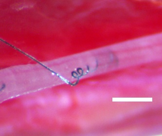
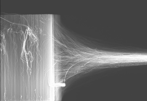
Chronic recording of vagal signals has been challenging due to the difficulty in recording high-quality signals in small autonomic nerves. Extraneural cuff electrodes have been effective, but they require desheathing of the nerve or have a low signal-to-noise ratio (SNR). Intraneural electrodes have higher SNR but are more invasive and have stability issues, particularly for small autonomic nerves. Carbon nanotube yarn (CNTY) electrodes have been shown to provide a stable, high- SNR interface for chronic recording in small autonomic nerves in rats, with high-quality signals continuing up to four months after implantation [1,2]. CNTY electrodes are a promising neural interface for interfacing with small peripheral nerves. These electrodes are small, low-impedance, and highly flexible biosensors, which makes them ideal for interfacing with small peripheral nerves. They are more than ten times more flexible than PtIr electrodes of the same diameter, making them highly attractive for use in various applications.

“Vagal tone” indicates overall levels of vagal activity
This technology has allowed for the first direct measurements of vagal tone in freely moving animals. [3] The vagus nerve is the largest autonomic nerve, innervating every organ in the body. “Vagal tone” is a clinical measure believed to indicate overall levels of vagal activity. Vagal tone is estimated indirectly through the heart rate variability (HRV), and abnormal vagal tone has been associated with many severe conditions such as diabetes, heart failure, and hypertension. However, vagal tone has never been recorded directly, leading to disagreements in its interpretation and influencing the effectiveness of vagal therapies. Using carbon nanotube yard electrodes, we were able to chronically record neural activity from the left cervical vagus in both anesthetized and unanesthetized rats. Here we show that tonic neural activity does not correlate with common HRV metrics with or without anesthesia. Additionally, we found that average vagal activity increases during inspiration compared to expiration; however, this vagal respiratory signal was also not correlated with HRV. This work is a significant step forward in recording technology and advancing our understanding of vagus nerve physiology and behavior.
The ability to continuously record spontaneous vagal-spiking activity from awake, freely moving rats for extended periods up to two weeks after implantation is a significant breakthrough in this field. Neural recording was synchronized with a continuous video recording of the subjects, and spike sorting was used to separate semi-distinct spike clusters correlated to animal behavior identified from the video recordings. Interspike interval distributions were found to change in response to food intake, presenting another neural feature that can be used to decode spontaneous vagal activity [4]. Several spike clusters show tuning to animal eating, and the firing dynamics of multiple decoded spike clusters can be used to classify eating compared to drinking, grooming, and resting behaviors.
Research focuses on using CNTY electrodes for chronic recording in small autonomic nerves
This line of research focuses on using CNTY electrodes for chronic recording in small autonomic nerves, such as the vagus and glossopharyngeal nerves, and for stimulation in larger somatic nerves and fascicles, such as the rat sciatic nerve. The study results demonstrate that CNTY electrodes can be used for the chronic recording small nerves, such as the vagus nerve, providing the first demonstration that spontaneous, physiologically relevant autonomic signals can be recorded chronically.
In what ways is the vagus nerve important?
The vagus nerve plays a vital role in homeostasis, reflex pathways, and responses to physiological changes. While individual vagal-spiking activity has been recorded from isolated fibers and acute intraneural recordings, this is the first-time neural spikes have been recorded within the vagus nerve in a chronic model. The study demonstrates that various spontaneous and physiologically relevant signals can be recorded from the rat vagus nerve from freely moving animals using CNTY electrodes.
Neural interfaces that allow for stable, high-SNR recordings are necessary for high-fidelity closed-loop control and chronic recording in animal models. The neurotechnology with Carbon Nanotubes Yarns has significant potential for vagus-nerve-stimulation therapy, which is a rapidly growing field with a wide variety of companies and studies investigating its use for the treatment of a wide variety of diseases, including epilepsy, obesity, and heart failure.
References
- McCallum GA, Xiaohong Sui, Chen Qiu, Joseph Marmerstein, Yang Zheng, Thomas E. Eggers, Chuangang Hu, Liming Dai, Dominique M. Durand, Chronic interfacing with the autonomic nervous system using carbon nanotube (CNT) yarn electrodes, Nature Scientific Reports, 1, 11723, 2017
- https://www.scipod.global/plugging-into-the-nervous-system-professor-dominique-durand-case-western-reserve-university/
- JosephT. Marmerstein, Grant A. McCallum, DominiqueM. Durand, Direct measurement of vagal tone in rats does not show correlation to HRV, ScienceReports, 1210 (2021)
- Joseph T. Marmerstein, Grant A. McCallum, Dominique M. Durand Decoding vagus nerve activity with carbon nanotube sensors in freely moving rodents, Biosensors- 1569057, 2022

This work is licensed under Creative Commons Attribution-NonCommercial-NoDerivatives 4.0 International.


