Adèle Ehongo explores the potential of optical coherence tomography for diagnosing peripapillary intra-choroidal cavitation in myopic eyes
Highly myopic complications, including Peripapillary Intra-Choroidal Cavitation (PICC), underspin the burden of myopia, a condition expected to affect almost half of the global population by 2050. (1)
EASY – Think about it and avoid anxiety and costly assessments
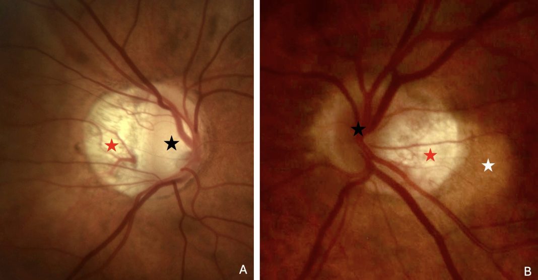
PICC is a circumscribed, yellow-orange lesion located at the outer border of the myopic conus (2) (the crescent-shaped atrophic area at the border of the optic nerve head (ONH)) (Figure 1). It may suggest a choroidal tumor, leading to unnecessary and anxiety-provoking assessments. (2) In up to 73.3% of cases, it is linked to visual field defects suggesting glaucoma, (3) especially since the yellow-orange appearance is absent in up to 53% of PICCs detected by Optical Coherence Tomography (OCT). (4)

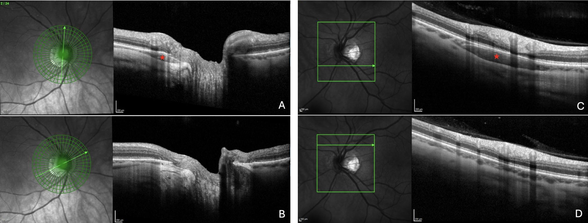
Two radial OCT slices of the same optic nerve head, with (A) and without (B) a peripapillary intrachoroidal cavitation (PICC). The PICC appears as a triangular hyporeflective intrachoroidal thickening (red star) with its base at the ONH.
Two linear OCT slices of the same eye with (C) and without (D) PICC. The PICC appears as a hyporeflective intrachoroidal thickening (red star) without modification of the plane of the anterior structures.
FAST – PICC diagnosis at your fingertip thanks to OCT
A quick scroll through the radial OCT slices of the ONH, provided ready for glaucoma assessment, reveals a hyporeflective triangular intrachoroidal thickening with its base at the ONH, which characterizes the PICC (5) (Figure 2A-B).
Furthermore, in case of doubt, linear OCT slices ahead from the ONH disclose the hyporeflective thickening, whose structures in front retain their plane, (5) confirming the diagnosis of PICC (Figure 2C-D).
By integrating a quick overview of the radial slices provided for glaucoma assessment into daily practice, the clinician improves his glaucoma diagnostic skills and avoids costly and worrisome examinations for the patient, highlighting OCT as the recommended tool for PICC screening. (6,7)
CLEAR – PICC is a suprachoroidal detachment
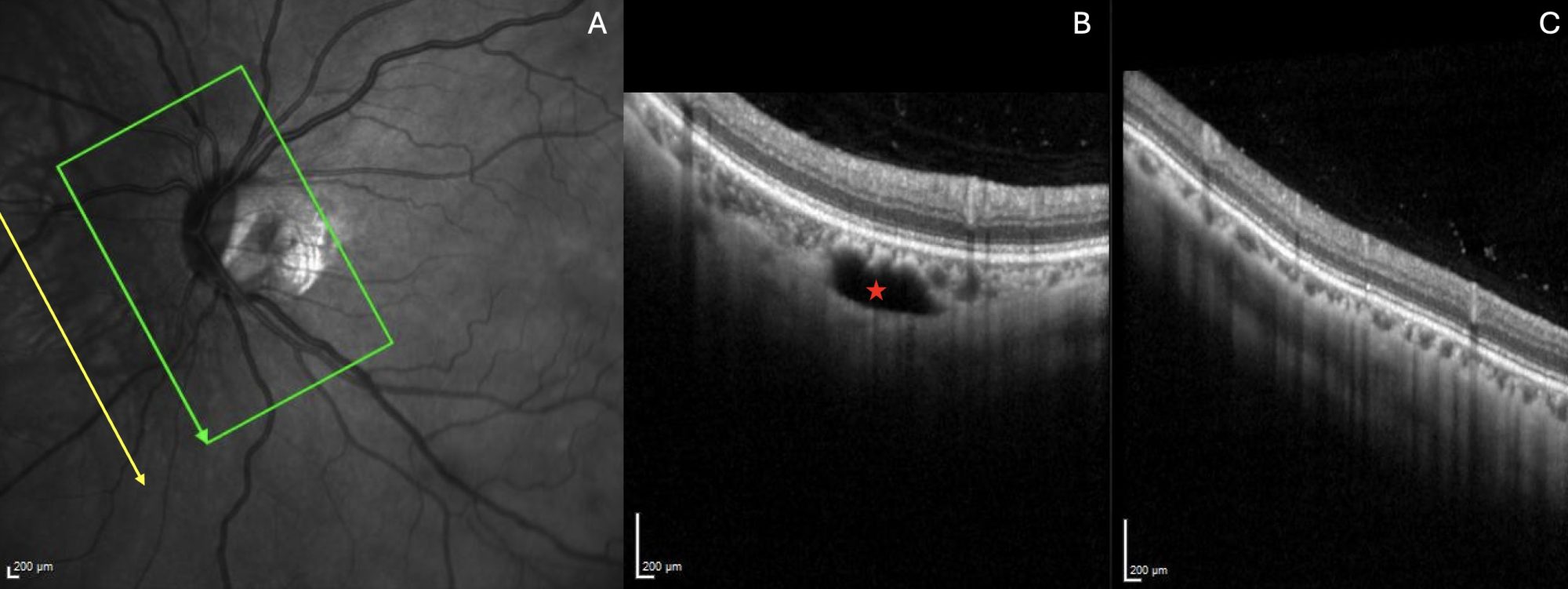
Still, ahead of the ONH and nasal to it, we recently documented that oblique OCT sections parallel to the major temporal blood vessels reveal the outer limit of the PICC where, thanks to the well-preserved appearance of the structures, the PICC is clearly revealed as a suprachoroidal detachment (6,7) (Figure 3).

Note: Reprinted from Analysis of Peripapillary Intrachoroidal Cavitation and Myopic Peripapillary Distortions in Polar Regions by Optical Coherence Tomography. Adèle Ehongo et al. ‘Clinical Ophthalmology 2022:16 2617-2629’ Originally published by and used with permission from Dove Medical Press Ltd.’
A CLUE – PICC and other myopic complications
How does this choroidal cleavage occur and why myopic eyes are more likely to develop PICC will be developed in a future edition, opening perspectives for understanding other myopic complications in order to mitigate the impact of this predicted myopic epidemic.
References
- Holden BA, Fricke TR, Wilson DA, Jong M, Naidoo KS, Sankaridurg P, Wong TY, Naduvilath TJ, Resnikoff S. Global Prevalence of Myopia and High Myopia and Temporal Trends from 2000 through 2050. Ophthalmology. 2016 May;123(5):1036-42. doi: 10.1016/j.ophtha.2016.01.006. Epub 2016 Feb 11. PMID: 26875007.
- Freund KB, Ciardella AP, Yannuzzi LA, Pece A, Goldbaum M, Kokame GT, Orlock D. Peripapillary detachment in pathologic myopia. Arch Ophthalmol. 2003 Feb;121(2):197-204. doi: 10.1001/ archopht.121.2.197. PMID: 12583785.
- Okuma S, Mizoue S, Ohashi Y. Visual field defects and changes in macular retinal ganglion cell complex thickness in eyes with intrachoroidal cavitation are similar to those in early glaucoma. Clin Ophthalmol. 2016 Jun 29;10:1217-22. doi: 10.2147/OPTH.S102130. PMID: 27418805; PMCID: PMC4935007.
- Dai Y, Jonas JB, Ling Z, Wang X, Sun X. Unilateral peripapillary intrachoroidal cavitation and optic disk rotation. Retina. 2015 Apr;35(4):655-9. doi: 10.1097/ IAE.0000000000000358. PMID: 25299968.
- Spaide RF, Akiba M, Ohno-Matsui K. Evaluation of peripapillary intrachoroidal cavitation with swept source and enhanced depth imaging optical coherence tomography. Retina. 2012 Jun;32(6):1037- 44. doi: 10.1097/IAE.0b013e318242b9c0.
PMID: 22466483. - Ehongo A, Bacq N. Peripapillary Intrachoroidal Cavitation. J Clin Med. 2023 Jul 16;12(14):4712. doi: 10.3390/jcm12144712. PMID: 37510829; PMCID: PMC10380777.
- Ehongo A, Bacq N, Kisma N, Dugauquier A, Alaoui Mhammedi Y, Coppens K, Bremer F, Leroy K. Analysis of Peripapillary Intrachoroidal Cavitation and Myopic Peripapillary Distortions in Polar Regions by Optical Coherence Tomography. Clin Ophthalmol. 2022 Aug 13;16:2617-2629. doi: 10.2147/OPTH.S376597. PMID: 35992567; PMCID: PMC9387167.


