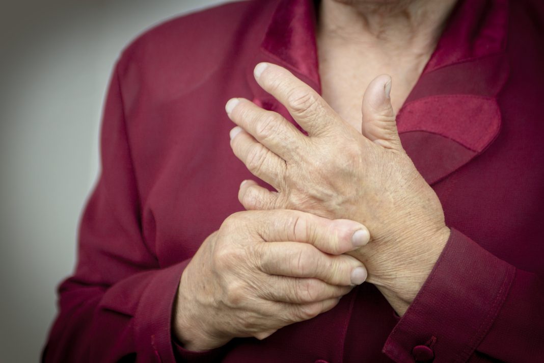
Osteoarthritis presents a significant societal and economic burden. Stina Simonsson from the University of Gothenburg explains how EU-funded projects are using 3D bioprinting to create functional cartilage for OA treatment
Osteoarthritis (OA) is a widespread condition that affects millions of people globally. As the population ages and the obesity epidemic continues, the incidence of OA is expected to increase. 3D bioprinting and stem cell therapies are heralded as the future of medical treatments for such conditions.
The global prevalence of OA
OA affects a significant portion of the Swedish population. Estimates suggest that about 7-8% of the population is diagnosed with OA, which translates to roughly 700,000 to 800,000 individuals. The condition is more common among older adults, with a higher prevalence observed in those over the age of 65.
OA is one of the most common forms of arthritis globally. According to World Health Organization (WHO) and other global health sources, approximately 10-15% of the global population over the age of 60 has some degree of OA. This means that about 100 million people over 60 worldwide are suffering from the condition, and 300 million people in total. These figures highlight the significant impact of OA on both national and global levels, emphasizing the importance of ongoing research, prevention, and treatment efforts.
RESTORE and autoCRAT
Two EU-funded projects, RESTORE and autoCRAT, aim to treat OA, for instance, by generating functional cartilage using 3D bioprinting technology, which is the focus of this article.
A Swedish research team within these EU projects has recently achieved a significant milestone towards novel OA treatment by successfully generating functional cartilage tissue through 3D bioprinting chondrocytes directly into OA lesions. This groundbreaking work was published in the journal CARTILAGE. (1) An earlier study by the same team, which demonstrated the formation of functional cartilage from 3D bioprinted induced pluripotent stem cells (iPSCs), was published in Scientific Reports. (2) These stem cells were derived by reprogramming the patient’s cells. (3) Utilizing iPSCs as a cell source for 3D bioprinting provides a foundation for developing personalized rejuvenated tissues or off-the-shelf products for clinical use. By achieving the required cell numbers, this approach could significantly reduce costs compared to traditional autologous cell treatments.
To rejuvenate a patient’s cells into pluripotent stem cells, or even into a new individual by cloning, was awarded the Nobel Prize in 2012. The cells require the expression of the transcription factor Oct4. (4-7) These cells can then theoretically multiply indefinitely and differentiate into any specialized cell type. Specialized cells are unable to multiply but are needed in their destined function.
Cartilage consists of specialized cells known as chondrocytes, which produce a collagen-rich extracellular matrix (ECM) containing proteoglycans and elastin fibers. There are three types of cartilage: elastic, fibrocartilage, and hyaline. These types differ in their collagen and proteoglycan content. Articular cartilage, a type of hyaline cartilage found in knee joints, is composed solely of chondrocyte cells, which play a crucial role in maintaining and repairing the ECM. Unlike mesenchymal stem cells (MSCs), chondrocytes are highly specialized and preferentially differentiate into cartilage rather than bone. Chondrocytes in tissue culture have been shown to dedifferentiate.
Autologous chondrocyte implantation (ACI), a procedure used for over 30 years to treat traumatic cartilage lesions, involves transplanting a patient’s own chondrocytes. (8) To generate mature articular cartilage via 3D bioprinting, the researchers used primary chondrocytes left over from ACI knee surgeries. Their experiments were designed with the understanding that a substantial number of cells are required to form tissue using 3D bioprinting. (2)
3D bioprinting technology allows for the combination of cells with biomaterials, enabling the creation of structures that match the shape of cartilage lesions or OA defects. Initially, an OA defect was scanned using MRI, CT, micro-CT, and a 3D scanner. The 3D scanner was the only device that accurately detected the lesion area. This 3D model served as a template for bioprinting primary chondrocytes to fit the exact shape of the lesion. The bioprinted chondrocytes formed cartilage tissue and produced collagen IIA isoform type B, which is specific to articular cartilage, as confirmed by two methods: RT-PCR and antibody staining. The levels of this collagen were higher in 3D bioprinted chondrocyte tissue compared to those cultured in 3D micromass, indicating that 3D bioprinting better supports cartilage formation.
Additionally, the bioprinted chondrocytes produced aggrecan (ACAN), a gene characteristic of articular cartilage. In contrast, chondrocytes cultured in a monolayer did not produce detectable levels of collagen II splice variant type B. This finding demonstrates that the bioink used in 3D bioprinting supports the production of mature articular cartilage, which could inform the design of next-generation scaffolds for treating articular cartilage injuries or OA. Within the RESTORE project, bioinks were developed, which could be combined with drugs to combat OA under the EU’s Horizon 2020 research and innovation program (grant agreement No 814558).
References
- B. Gatenholm, C. Lindahl, M. Brittberg and S. Simonsson, ‘Collagen 2A Type B Induction after 3D Bioprinting Chondrocytes In Situ into Osteoarthritic Chondral Tibial Lesion’, Cartilage: (2020), p. 1947603520903788.
- D. Nguyen, D. A. Hagg, A. Forsman, J. Ekholm, P. Nimkingratana, C. Brantsing, T. Kalogeropoulos, S. Zaunz, S. Concaro, M. Brittberg, A. Lindahl, P. Gatenholm, A. Enejder and S. Simonsson, ‘Cartilage Tissue Engineering by the 3D Bioprinting of iPS Cells in a Nanocellulose/Alginate Bioink’, Sci Rep, 7: 1 (2017), p. 658.
- C. Borestrom, S. Simonsson, L. Enochson, N. Bigdeli, C. Brantsing, C. Ellerstrom, J. Hyllner and A. Lindahl, ‘Footprint-free human induced pluripotent stem cells from articular cartilage with redifferentiation capacity: a first step toward a clinical-grade cell source,’ Stem Cells Transl Med, 3: 4 (2014), pp. 433-47.
- Kazutoshi Takahashi, Koji Tanabe, Mari Ohnuki, Megumi Narita, Tomoko Ichisaka, Kiichiro Tomoda and Shinya Yamanaka, ‘Induction of pluripotent stem cells from adult human fibroblasts by defined factors’, Cell, 131: 5 (2007), pp. 861-72.
- J. A. Byrne, S. Simonsson, P. S. Western and J. B. Gurdon, ‘Nuclei of adult mammalian somatic cells are directly reprogrammed to oct-4 stem cell gene expression by amphibian oocytes’, Curr Biol, 13: 14 (2003), pp. 1206-13.
- J. B. Gurdon, T. R. Elsdale and M. Fischberg, ‘Sexually mature individuals of Xenopus laevis from the transplantation of single somatic nuclei,’ Nature, 182: 4627 (1958), pp. 64-5.
- K. Takahashi and S. Yamanaka, ‘Induction of pluripotent stem cells from mouse embryonic and adult fibroblast cultures by defined factors’, Cell, 126: 4 (2006), pp. 663-76.
- M. Brittberg, A. Lindahl, A. Nilsson, C. Ohlsson, O. Isaksson and L. Peterson, ‘Treatment of deep cartilage defects in the knee with autologous chondrocyte transplantation,’ N Engl J Med, 331: 14 (1994), pp. 889-95.
- Tomi Kalpio Sanna Turunen, Carl Lindahl, Chirag Jain Mysore Shanthinathan, Tahsin Akhter, Sebastian Concaro, Stina Simonsson ed., Chapter 12 – Future solutions for osteoarthritis using 3D bioprinting of articular cartilage, 2023), 335-69.
- H. Evenbratt, L. Andreasson, V. Bicknell, M. Brittberg, R. Mobini and S. Simonsson, ‘Insights into the present and future of cartilage regeneration and joint repair’, Cell Regen, 11: 1 (2022), p. 3.
For more information(9,10) about the RESTORE or autoCRAT projects and its partners, visit: https://restoreproject.eu or https://autocratproject.eu

This work is licensed under Creative Commons Attribution-NonCommercial-NoDerivatives 4.0 International.


