Advancements in dental imaging over the past two decades have been remarkable, such as cross-polarization OCT; Yihua Zhu and his team at the University of California, San Francisco, have been investigating different diagnostic imaging methods
Yihua Zhu and his team at the University of California have extensively explored innovative imaging techniques, comparing them with conventional methods. Their focus has been on enhancing short wavelength infrared (SWIR) technologies, developing a dual probe for investigating dental cavities, and, more recently, introducing a novel SWIR multispectral transillumination and reflective imaging method. This research signifies a significant leap in providing a highly sensitive and efficient approach to visualizing dental cavities, tracking tooth decay progression, and assessing prevalent dental issues.
Among these issues, dental cavities or caries resulting from bacterial-induced mineral loss pose a frequent challenge in dentistry. An ongoing issue has been the lack of standardized diagnostic methods for cavities. Until recently, accurate assessment tools for cavities and other dental conditions have been inadequate. Visible cavities such as occlusal and root caries were typically diagnosed through visual inspection and tactile examination, methods prone to subjectivity and imprecision.
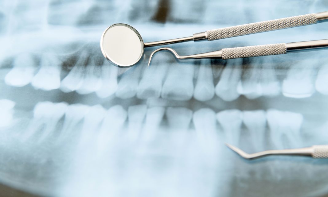
While X-rays have conventionally served diagnostic purposes in oral health, they have limitations, especially in assessing cavity depth. This often leaves dental professionals relying on estimations based on visual evidence and their experience, potentially leading to unnecessary dental procedures.
Expanding dental imaging research
Over the last two decades, a dedicated group led by Yihua Zhu at the University of California’s Department of Preventive and Restorative Dental Sciences has been pioneering new dental tools. Zhu’s contributions have expanded on existing dental imaging research, particularly in short wavelength infrared (SWIR) reflectance. Their introduction of dual techniques, combining transillumination imaging with a newly developed probe, has significantly advanced these methods.
While these methods are typically used independently, Zhu and his mentor, Professor Daniel Fried, have innovatively combined both modalities. Their recent focus has involved fine-tuning this amalgamated tool by exploring SWIR using various light wavelengths and comparing outcomes with standard dental methods. Additionally, Zhu and his team have delved into the significance of cross-polarization optical coherence tomography (CP–OCT) in diagnosing dental caries.
Traditionally, dental diagnoses have relied on a combination of X-rays and visual assessments, offering valuable insights widely used in dental practice. However, their effectiveness is limited, especially in evaluating root and occlusal caries. X-rays can only detect mineral loss between 30–60%, missing early-stage caries. The visual assessment also falls short, struggling to distinguish between non-carious staining and carious lesions, leading to potential overtreatment concerns.
Statistics suggest that about a third of regular dental patients might have suspicious lesions, like cracks or cavities, typically in posterior teeth. Dental practitioners urgently require new methods to gauge cavity depth and assess whether decay has reached the tooth’s dentin—the inner layer beneath enamel protecting nerves. Precise evaluation in this area is crucial as issues here may necessitate surgery.
Short-wave infrared (SWIR) and near-infrared (NIR) imaging
Around two decades ago, innovative imaging techniques like short-wave infrared (SWIR) and near-infrared (NIR) imaging emerged. SWIR light allows transparent enamel imaging, enabling occlusal transillumination—capturing images of the tooth from its biting surface or below the gum line. This method proved promising for early caries detection and assessing cavity severity without ionizing radiation. SWIR imaging efficiently detects early demineralization caused by plaque, allowing intervention before severe damage, often treatable conservatively with fluoride varnish to promote remineralization.
Combining different modalities became a key research focus due to the strengths and limitations of each method. While single-wavelength SWIR imaging offers high sensitivity, it poses challenges with false positives like glares and hypomineralization, potentially mistaken for dental decay.
CP-OCT: A non-invasive dental imaging technique
Mr Zhu and his team have recently explored CP-OCT, a non-invasive imaging technique utilizing polarized light to generate 3D tooth images. This method enhances image contrast, allowing for detailed visualization of the tooth’s cross-sectional anatomy. CP-OCT has demonstrated utility in identifying root and coronal caries, cementum loss, and severe demineralization at the root surface— issues typically concealed from view.
The exceptional resolution of CP-OCT enables the identification of the transparent remineralized surface layer that forms atop dental caries. This layer signifies the tooth’s natural healing process, like scabs on skin wounds. Detecting this remineralized layer indicates the lesion’s initiation of self-repair, indicating that further treatment may not be necessary. However, this layer is invisible under visible light and too thin for detection through radiographs. Only through histological examination of extracted teeth can its presence be confirmed.
Utilizing CP-OCT, Mr Zhu and his team successfully identified this remineralized layer in living subjects and monitored its growth over time. This breakthrough can potentially revolutionize conservative dental treatments by identifying ‘arrested’ caries, eliminating the need for mechanical intervention. These advancements made by Mr Zhu’s research group mark a substantial leap in modern dentistry, particularly dental diagnostics.

This work is licensed under Creative Commons Attribution-NonCommercial-NoDerivatives 4.0 International.


