Anders Brostrøm & Kristian Mølhave from DTU Nanolab, Technical University of Denmark, share their views on improving the foundation for assessing the health impact of particles and aerosols
Aerosol particles, also known as Particulate Matter (PM), have a major influence on our lives.
In our local ambient, indoor and workplace environment, the particles emitted from industry, agriculture, transport and household activities, can cause severe health effects. Today PM is regulated based on mass concentration, though it is well accepted that particle mass is insufficient for assessing risk on its own, as the fate and influence of PM on human health depend on their physicochemical properties, for example, size, shape, and composition.


These properties can, however, vary individually between particles and are difficult or not possible to measure using standard techniques, bringing a need for new PM characterisation tools. We explore a new method to characterise PM by electron microscopy, which can offer detailed physicochemical information on a single particle basis, thus, providing a better foundation for assessing risk from PM exposure.
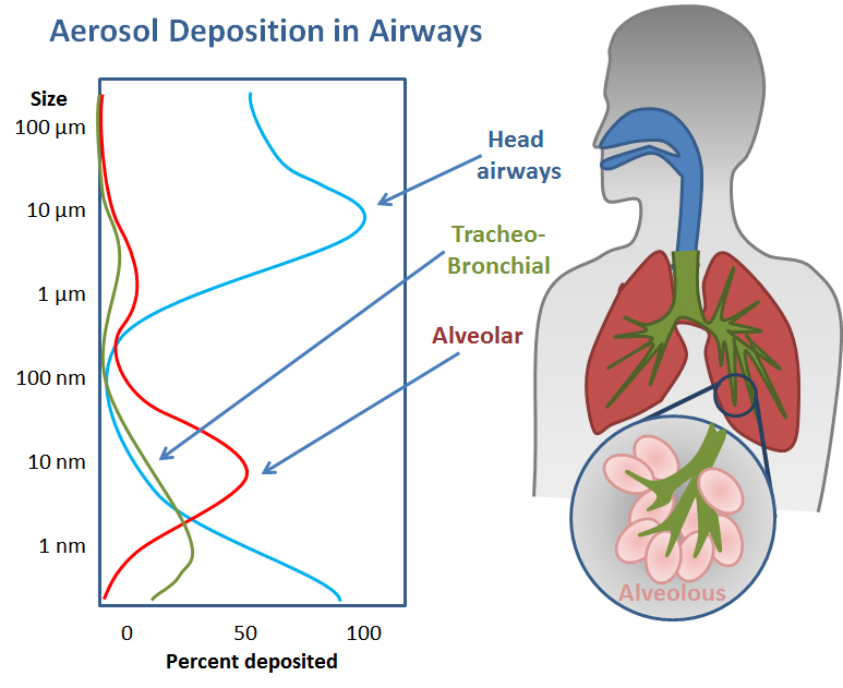
The fate and role of airborne particles when inhaled are governed by their physicochemical properties. The particle size determines where in the respiratory system they are most likely to deposit, as shown in Figure 1, and strongly influence the health effect of the exposure. Particles vary individually in size, shape, composition and physical state, and also over time due to evaporation and condensation processes in the ever-changing
environment.
Particulate matter (PM) exposure assessments are often given based on real- time number or mass concentration measurements. This strategy is sufficient in scenarios where only a single type of PM is present – but this is rarely the case. Ambient, indoor, and workplace environments often contain aerosols of much higher complexity, consisting of a mixture of several particle types e.g. soot from engines, salts from atmospheric sea spray, soil and dust particles, or specific particles from industrial production. The particle types will likely have significantly different properties and, thus, pose different risks to human health. To assess the health risk of complex aerosols, the relative contribution of each particle type to the overall aerosol population must be measured.
Several kinds of aerosol measurement instruments are available, but most bring no information on the shape or composition of individual particles and, therefore, cannot distinguish between particle types. In addition, most traditional measurement
techniques do not function well for non-spherical particles, which include high-risk particles such as fibers.
“The fate and role of airborne particles when inhaled are governed by their physicochemical properties. The particle size determines where in the respiratory system they are
most likely to deposit, and strongly influence the health effect of the exposure.”
Scanning electron microscopy (SEM) can provide images of particles collected onto a substrate. Individual particles can be distinguished from the images, allowing measurements of their physical size and shape. In addition, SEM can be coupled with energy-dispersive X-ray spectroscopy (EDS) that can give information on the elemental composition of each particle. The method, therefore, has the potential to bring information on many of the relevant particle properties and could be a vital supplement to real-time measurements.

We studied if SEM/EDS analysis of complex aerosols allows for automatic identification and separation of particles in an aerosol population and whether it can be used to classify them individually, based on their physicochemical properties, even for nano-sized particles.
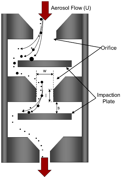
The applied analysis consists of several steps.1 The first step is to collect particles onto a smooth surface, which can be brought to the microscope for analysis. This was done via impaction (Figure 2), which can collect aerosol particles directly onto SEM suited substrates, using a small transportable impactor and pump.
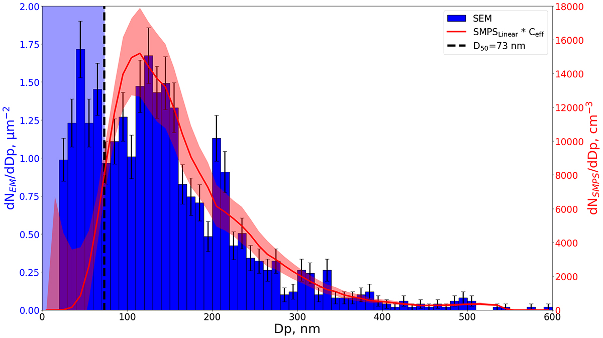
The method showed good agreement when comparing size distribution results to the well-established Scanning Mobility Particle Sizer (SMPS), as seen in Figure 3.
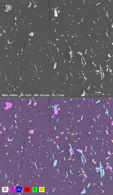
To push the SEM method to the limits, another test was performed using a complex aerosol, consisting of particles with significantly different physicochemical properties: salt particles (NaCl), soot-like carbon-based particles (Printex90), and pristine Al/Si fibers.2 An example of SEM/EDS images from the experiments are shown in Figure 4.

The EDS results made it possible to distinguish and categorise individual particles, based on their elemental composition: NaCl particles appear orange (red Na combined with yellow Cl), the Al/Si fibers appear cyan (blue Al combined with green Si), while Printex90 particles are magenta as they only consist of carbon. As a result, the relative contribution of each particle type to the overall aerosol population could be estimated. In addition, the geometry, for example, the size and shape of each particle were determined by automated image analysis of the SEM images, enabling particle type-specific analysis.
Overall, the SEM/EDS analysis made it possible to distinguish fibrous particles from particles with other shapes, as well as to distinguish and classify particles by their composition (NaCl, Printex90 and Al/Si fibers). This enabled estimates of the relative
contribution of each particle class to the overall aerosol population, which proved viable for particles as small as 50 nm.
In summary, we showed that SEM/EDS analysis enables a more detailed physicochemical characterisation of PM compared to other aerosol instruments. The method can provide detailed individual particle and aerosol information on size, shape and elemental composition, which are crucial information for improving the current foundation for PM exposure and risk assessments. Also, the analysis can facilitate source identification based on identified particle types, which is essential for developing efficient preventive strategies.
References
1 Brostrøm, A. et al. Improving the foundation for particulate matter risk assessment by individual nanoparticle statistics from electron microscopy analysis. Sci. Rep. 9, 8093 (2019).
2 Brostrøm, A., Kling, K. I., Hougaard, K. S. & Mølhave, K. Complex Aerosol Characterization by Scanning Electron Microscopy Coupled with Energy Dispersive X-ray Spectroscopy. Submitt. to Sci. Reports (2019).
We gratefully acknowledge funding from the Danish NanoSafety Centre and The National Research Center for the Working Environment (NFA) in Denmark.
*Please note: This is a commercial profile


