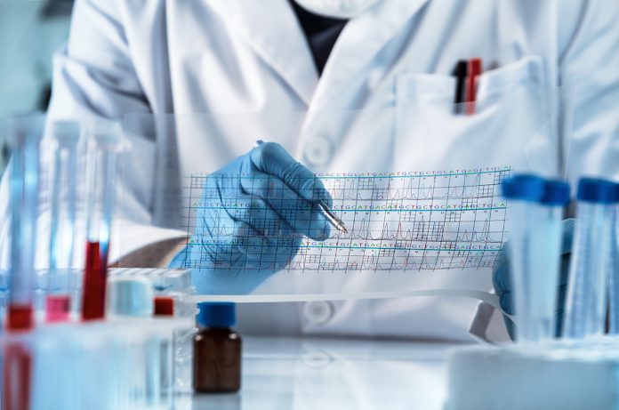
Richie Kohman, Synthetic Biology Platform Lead at Wyss Institute at Harvard, explains the use of next-generation sequencing to analyse biological tissues in a spatially resolved context
Next-generation sequencing involves the massively parallel readout of DNA sequences, usually accomplished in commercial instruments. The cost per base of sequencing has dropped over one million-fold over the past two decades resulting in a revolution in how biological research is performed. This amazing progress is in part due to engineering advances of the sequencing hardware as well as in the chemistry within the instruments. The speed, cost, and information content associated with DNA sequencing have now incentivised researchers to use NGS to analyse other molecules of interest, such as RNA, by converting them to DNA.
There is now increasing interest in harnessing the power of NGS to analyse biological tissue. Tissues are immensely complex and contain an ocean of molecules that determine their function. Researchers are interested in interrogating these tissues to both understand fundamental insight about the structure as well as to understand disease and create valuable diagnostic or therapeutics.
The earliest examples of using NGS to analyse tissue involve the homogenization of the samples to liberate molecules of interest, usually RNA. These “bulk sequencing” approaches provide a rapid way to directly access the contents of the tissues. The simplicity of this approach, however, comes at the expense of cellular resolution – tissues are not homogenous but rather contain a variety of different cell types and bulk approaches cannot resolve this level of detail. To alleviate this issue, researchers developed a suite of techniques that can separate dissociated tissues into individual cells before analysis. These single-cell sequencing techniques have emerged as powerful methods that rapidly profile entire populations of cells.
Despite the popularity of bulk or single-cell sequencing of tissues, both of these approaches suffer from a loss of spatial context. Not only are tissues cellularly heterogeneous, but they also exhibit spatial intricacies which are crucial to the function of the organ. Dissociation of the samples eliminates this spatial record of molecules being analysed. A new research field has now emerged to analyse tissue with regard to spatial context. This article will provide a brief overview of these technologies. In most of these cases, the targets of interest for researchers are not genomic DNA but rather a different molecule that can be investigated using the NGS workflow or chemistry. Each of these categories will be elaborated upon in future articles dedicated to these topics.
Dissection
Perhaps the most straightforward way towards retaining spatial information from tissues is to utilise the existing methods for bulk tissue sequencing but modify the portion of the tissue that is analysed. A variety of dissection-based approaches use this strategy. In laser capture microdissection (LCM) and GeoSeq, a low-power infrared laser beam is irradiated onto histological sections to remove only a desired portion onto a film for further processing and sequencing. In a technique called RNA tomography or tomo-seq, three identical samples are sectioned in perpendicular planes and subject to bulk sequencing. The location of transcripts of interest are determined by using the overlapping data to perform a 3D reconstruction. In ProximID, samples are partially dissociated into clusters of cells that are sequenced. The adjacent associations can then be determined from the data. The major limitation in all of these approaches is throughput as the dissection process is rate-limiting.
Capture
A suite of techniques has been developed that involve a transfer or capture of targets off of the tissue location. Following capture, the molecules can be analysed by NGS after converting them to DNA. As with many of these approaches, they are focused on the RNA analysis because of the usefulness of spatial gene expression. Techniques such as Spatial Transcriptomics, 10X Visium, High-Definition Spatial Transcriptomics (HDST), and Slide-seq all involve the pulldown of RNA from the tissue slices onto surfaces in a way urface that retains the spatial location of the transcript. An alternative approach has been demonstrated by Nanostring’s GeoMx approach which allows the removal of targets only at the area or UV light irradiation. The approach is notable in that, in addition to RNA, proteins can be queried spatially. This marks one of the unique spatial NGS techniques that has expanded beyond transcriptomics. This advantage has high throughput but is limited by the spatial resolution of the capture step.
In situ
The third approach involves fully in situ approaches. Possibly the most technically complicated category, these methods analyse the targets of interest directly on the biological sample. Many of these approaches involve innovations in the initial chemistry that prepares the tissue for sequencing. Fluorescent in situ sequencing (FISSEQ), in situ sequencing (ISS) using padlock probes, and Spatially-resolved transcript amplicon readout mapping (STARmap) all differ in their initial steps to anchor and convert molecules of interest into a form that can be sequenced, however, the subsequent steps are similar. Despite the challenges of performing all steps within the biological sample, this category shows the most diversity concerning the type of targets profiled. In addition to RNA, there have been advances allowing the in situ analysis of genomic DNA as well as proteins. The major advantage of this category is the high spatial resolution which is limited only by microscopy optics.
Analysis of synthetic biology signatures
The final category is less about how NGS is used to analyse the tissue sample and more about what role the molecules being analysed are playing. Up until now, all of the methods discussed have analysed natural, endogenous molecules within the sample. However, there is a new trend emerging that uses synthetic biology to engineer biological samples to express targets of interest at specific times or locations such that they can be ready out at a later time.
Phenomena that can be elucidated by this approach include cellular development or brain connectivity. In these applications there lacks a convenient marker that can be traced to simplify the investigation; therefore, synthetic biologists are developing novel means to position these within tissues.
*Please note: this is a commercial profile


