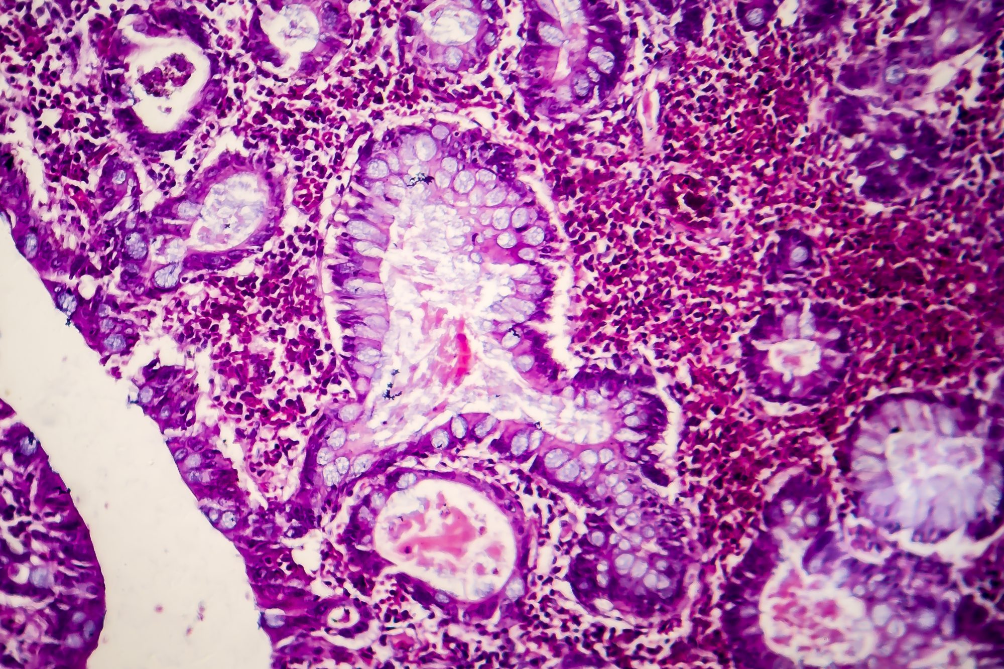
Mr Sanj Lallie, Commercial Director of Digital Pathology from Source LDPath, describes the pathway for a nationwide uptake of digital pathology and Al to build the future of medicine
For almost 10 years of providing specialist histopathology services to the NHS, LDPath (now Source LDPath, “Source LDP”*) has serviced the increased demand for specialist histopathology expertise. The growing workload in UK-based histopathology departments and a paucity of histopathologists is now a palpable crisis.
These supply and demand difficulties have resulted in huge backlogs and thus, often breached NICE cancer guidelines. Worryingly, it is projected that the lack of adequate specialists in the field of histopathology will last at least ten years. There are currently no plans published by any health authority within the UK on how to combat this.
A shortage of pathologists coupled with a growing incidence of cancer creates a strong foundation for necessity – and necessity is the mother of invention. There are other forces at play – human and economic costs, particularly of diagnostic errors. All of this creates a fertile environment for leveraging new technologies, especially that of AI.
It is well known that the histopathological diagnosis can have considerable inter-reader variability. For example, a 75% overall concordance rate in the evaluation of breast biopsies and an inter-observer agreement of only 48% for a diagnosis of atypical hyperplasia has been reported (Elmore et. al. 2015). Considerable discordance has been repeatedly documented in the interpretation of melanocytic neoplasms on skin biopsies (Elmore et.al. 2017). Such findings clearly indicate the salience of limited precision and interpretive accuracy.
AI methodologies aim to address difficulties within pathology
AI methodologies within pathology aim to address these difficulties, building upon the data-driven revolution and digitisation of workflows, and indeed there are nationwide programmes to develop the uptake, development and validation of AI.
AI already offers a smorgasbord of support to pathologists and has been successfully implemented in the Nordic regions. It carries out laborious, subjective, and repetitive tasks arriving at conclusions based on statistical probability and presenting its findings to the supervising Consultant for approval or rejection. From IHC scoring, tumour location, mitotic counting and margin location, dynamic, clinical-grade tools are readily available to save consultants’ time and deliver improved accuracy. Augmenting diagnosis of the screening programmes can reduce workloads by 70-85% and huge potential time savings, particularly in lymph node screening and prostate screening.
As AI matures, it takes on increasingly difficult tasks with precision unmatched by the human eye, but in a role that augments rather than replaces — a friendly disrupter to traditional workflows — allowing experts to deploy their expertise in new ways. As digital drivers push ahead with new technologies, as data accumulate and geographical restraints for working are removed, histopathology evolves into a more dynamic state.
Gone are the days when ‘a microscope required a table lamp as its external light source and a mirror on its base to make sense of histology and cytology slides’ (Laszlo Igali, Ferenc Igali, 2019). Pathology departments across the country, and indeed across the world, are connecting to their teams and each other – and crucially with data – in global digital workflows.
The use of Digital Slide Scanners, which automate the digitisation of biopsy slides, is now widespread and laboratories across the country are scanning slides at an industrial rate. However, fewer pathologists, extreme pressures of demand and legacy, hard-to-change systems provide significant barriers in allowing these innovative technologies to benefit service users and improve outcomes. Simply ‘going digital’ and scanning slides is only the first step in the digital revolution.
‘We are now entering a new era of augmented intelligence (AuI), whereby AI will work alongside clinicians in the decision-making process’
(Laszlo Igali, Ferenc Igali, 2019)
Over time, digital workflows will become ubiquitous, and Source LDP can help bridge the gaps and help partners leverage new digital capabilities.
Source LDP has a history of innovation and flexibility in adapting and opening up new digital possibilities for partners as they take their first, or further, steps into the digital world, including assisting with AI. Source LDP has worked with Ibex, a developer of high precision diagnostic tools, to provide algorithms that can be employed for clinical practice in a user-friendly way to facilitate the digital workflow within the histopathological landscape.
By providing validated apps, via the Source LDP ecosystem, individual NHS organisations have no need to build costly infrastructure, allowing them to focus on implementation to experience the benefits of these augmented digital workflows.

Digital capabilities reduce pressure on NHS histopathology departments
Using the fast turnaround services while exploring new digital capabilities allow NHS histopathology departments to relieve pressure and to free up crucial staff time to deploy their expertise in a more targeted and effective manner.
Through the development of proprietary software and working with others at the forefront of the revolution such as Ibex, Source LDP provides a litany of benefits – solutions that save time, improve testing accuracy and reproducibility in laboratories – while delivering results faster, and at a lower cost.
Just over the horizon lie new waves of technology – simplified, context-driven and personally customisable digital workflows with seamless human-computer interaction with many other advancements, such as 3D visualisation and scanning, including live volumetric measurements, remote holographic assistance and myriad other innovative ideas in VR/AR, as well as increasingly powerful augmented intelligence. Embracing validated and tested new technologies will lead to improvements for every stakeholder and everyone impacted by the workflow and greatly increase the effectiveness of each pathologist and department by giving them new tools with which to build the future of medicine.
Source LDP aspires to make these available to every institution using digital pathology with whom they will actively collaborate to accelerate their own digital journey.
Learn more about Source LDP
Source LDP offers a unique UK-based comprehensive rapid digital cellular pathology reporting service utilising state-of-the-art histology whole slide imaging capabilities integrated with a pathologist designed Lab Information Management System.
Founded and led by NHS consultant histopathologists, Source LDP’s reporting capability has been enabled by bringing together the largest digital network of specialists in the UK. Over 450 hospitals, clinics and clinicians across the country now benefit from Source LDP’s unrivalled specialist reporting services.
As pioneers of digital pathology, Source LDP offers:
- End-to-end Digital Reporting
- A managed histopathology service, from courier to report delivery.
- Unrivalled turnaround times.
- Specialist reporting in every
- An SSL encrypted LIMS application enabling you to access your reports quickly and safely.
- Multi-Disciplinary Meeting support.
- A unique, integrated second opinion feature, providing instant access to national and international expertise.
- AI quality assurance tools.
References
- Elmore JG, Longton GM, Carney PA, et al. Diagnostic Concordance Among Pathologists Interpreting Breast Biopsy Specimens. JAMA. 2015;313(11):1122–1132. doi:10.1001/jama.2015.1405.
- Elmore JG, Barnhill RL, Elder DE, Longton GM, Pepe MS, Reisch LM, Carney PA, Titus LJ, Nelson HD, Onega T, Tosteson ANA, Weinstock MA, Knezevich SR, Piepkorn MW. Pathologists’ diagnosis of invasive melanoma and melanocytic proliferations: observer accuracy and reproducibility study. BMJ. 2017 Jun 28;357:j2813. doi: 10.1136/bmj.j2813. Erratum in: BMJ. 2017 Aug 8;358:j3798. PMID: 28659278; PMCID: PMC5485913.
- Ehteshami Bejnordi B, Veta M, Johannes van Diest P, et al. Diagnostic Assessment of Deep Learning Algorithms for Detection of Lymph Node Metastases in Women With Breast Cancer. JAMA. 2017;318(22):2199–2210. doi:10.1001/jama.2017.14585.
- Campanella, G., Hanna, M.G., Geneslaw, L. et al. Clinical-grade computational pathology using weakly supervised deep learning on whole slide images. Nat Med 25, 1301–1309 (2019). https://doi.org/10.1038/s41591-019-0508-1
- Snead DR, Tsang YW, Meskiri A, Kimani PK, Crossman R, Rajpoot NM, Blessing E, Chen K, Gopalakrishnan K, Matthews P, Momtahan N, Read-Jones S, Sah S, Simmons E, Sinha B, Suortamo S, Yeo Y, El Daly H, Cree IA. Validation of digital pathology imaging for primary histopathological diagnosis. Histopathology. 2016 Jun;68(7):1063-72. doi: 10.1111/his.12879. Epub 2015 Dec 6. PMID: 26409165.
- Igali, (2019) The Road to Augmented Pathology, Available at: https://thepathologist.com/inside-the-lab/the-road-to-augmented-pathology (Accessed February 2022).
*LDPath Ltd is now Source LDPath following the acquisition by SourceBio International plc (AIM:SBI) in March 2022.
Please note: This is a commercial profile
© 2019. This work is licensed under CC-BY-NC-ND.


