AI is beginning to find its place within the healthcare sector, particularly in medical imaging and tumour detection
Imaging is one of the most promising areas in detecting and analysing tumours; learning algorithms are now being developed to identify the location, size, and type of tumours from complex medical scans.
Recent developments, highlighted by the success of the Karlsruhe Institute of Technology (KIT) team in the international auto PET competition, show how these AI-driven tools are becoming increasingly effective at supporting doctors in diagnosing and treating cancer.
The importance of medical imaging in cancer diagnosis
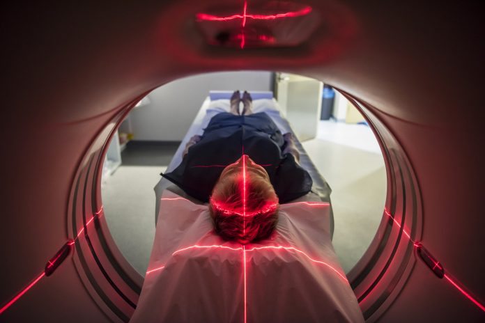
Accurate imaging plays an important role in diagnosing cancer and guiding treatment decisions. Two key techniques are positron emission tomography (PET) and computed tomography (CT).
PET scans offer insights into the metabolic activity of tumours by using a radioactive substance, typically a glucose analogue, which tumours absorb at higher rates than normal tissues.
CT scans, on the other hand, provide detailed images of the body’s internal structures, helping doctors pinpoint the exact location and size of tumors.
While these technologies have revolutionised cancer diagnosis, analysing the resulting images remains a time-consuming and intense task. A single patient may have dozens or even hundreds of tumours that need to be identified, measured, and tracked over time. Currently, radiologists and oncologists manually evaluate 2D slice images to mark the location of each tumor, a process that can take hours for each patient.
AI has the potential to save time and improve accuracy
This is where AI can be used. Using machine learning algorithms in medical imaging aims to automate and expedite this process, allowing healthcare professionals to focus more on interpreting results and making treatment decisions rather than spending time on repetitive manual tasks.
AI can help quickly identify and segment tumor lesions, ensuring no pathology is overlooked and improving the accuracy of the overall evaluation.
One of the standout teams in this effort was from the KIT’s Computer Vision for Human-Computer Interaction Lab (cv:hci). In 2022, they placed fifth in the autoPET competition, a global challenge that focused on automating the segmentation of tumor lesions in PET/CT scans.
The competition was organised by leading medical research institutions, including Tübingen University Hospital and LMU Hospital Munich, and 27 teams worldwide participated. The challenge was developing algorithms to identify and segment metabolically active tumor lesions from a large set of annotated PET/CT datasets.
Advances in deep learning algorithms
The top-performing teams in the competition developed algorithms that use deep learning techniques and advanced machine learning methods that utilise multi-layered neural networks to process large amounts of data and recognise complex patterns.
These algorithms could automatically detect and segment tumor lesions with impressive precision.
In collaboration with experts from the IKIM Institute for Artificial Intelligence in Medicine, the research team from KIT contributed to an important finding: an ensemble approach, which combines several algorithms, performed better than any single algorithm alone.
By merging the strengths of different AI models, the ensemble method was able to more accurately and efficiently identify tumor lesions, significantly enhancing the overall performance of the AI system.
Fully automated analysis
Although these AI models have shown impressive results, work still needs to be done. The quality and quantity of the data used to train the algorithms are important, and further research is needed to improve their robustness. For example, AI models must be able to handle variations in image quality, patient conditions, and the specific types of tumors being scanned. The long-term goal is to achieve a system capable of fully automating the analysis of PET and CT scans, allowing doctors to make faster, more reliable decisions regarding cancer treatment.


