Ductal carcinoma in situ (DCIS) is an early form of breast cancer that can be treated with surgery. Surgery can involve removal of the entire breast or partial removal of the tumour and the breast tissue surrounding it. In the case of partial removal, the challenge is knowing how much to remove. This is important because leaving any tumour behind is a major risk factor for tumour recurrence. It has been reported that, in 38-72% of all cases, the surgery fails to remove all of the affected tissue, highlighting a major health problem for women. Our approach to this problem has been to develop a predictive mathematical model that enables surgeons to anticipate the size of DCIS tumours and plan the surgery in advance to avoid leaving any tumour behind.
We call this approach mathematical pathology because all data being supplied to the mathematical model can be derived from a patient’s biopsy, which is used to make the diagnosis. In our recent modelling work on DCIS (Edgerton et al., Anal Cell Pathol 2011, PMC3613121), we sought to determine if an accurate prediction of tumour size is achievable through measurements that are taken from a single biopsy. The key parameters of the model are the distance for nutrients to diffuse into the tumour, the ratio of apoptosis to proliferation rates in the tumour, and the tumour size. The apoptosis to proliferation rate and diffusion distance can both be measured easily from a biopsy specimen, allowing one to solve for the tumour size. This is described in more detail in the Box.
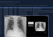
Our data show that surgical volumes determined by radiology and tumour grade are largely inaccurate when compared to surgical results. We examined 17 excised DCIS tumours and found that mammography overestimated the tumour size in ten cases and underestimated the tumour size in seven cases. This indicates that the correlation between mammography, nuclear grade, and the final observed tumour size is poor. However, the correlation between the sizes observed after surgery and those predicted by the model were close.
The clinical implication of our model predictions is that with standard mammogram and pathologic specimens, physicians should be able to accurately predict the surgical volume of DCIS tumours mathematically. This study represents a proof of principle that it is possible to incorporate a mathematical modelling step within the current clinical practice to aid in and improve surgical planning by estimating the surgical volume and the outcome of surgery before treatment. The practice will lead to less subjective analysis of tumours and an individualized approach to surgical removal of DCIS.
Additional resources:
www.internationalinnovation.com/taking-cancer-out-of-the-equation/
www.pnas.org/content/110/35/14266.long
www.hindawi.com/journals/acp/2011/803816/abs/
www.sciencedirect.com/science/article/pii/S0022519312000665
Eugene J. Koay1, Vittorio Cristini2,3, and Zhihui Wang2,3
1 Department of Radiation Oncology, University of Texas MD Anderson Cancer Center, Houston, TX 77030, USA
2 Department of NanoMedicine and Biomedical Engineering, University of Texas Health Science Center at Houston (UTHealth) McGovern Medical School, Houston, TX 77054, USA
3 Brown Foundation Institute of Molecular Medicine, University of Texas Health Science Center at Houston (UTHealth) McGovern Medical School, Houston, TX 77030, USA
Vittorio Cristini, Ph.D.
UTHealth McGovern Medical School, Department of Nanomedicine and Biomedical Engineering
Center for Advanced Biomedical Imaging (CABI)
Tel: +1 713 486 2315
Fax: +1 713 796 9697
Vittorio.Cristini@uth.tmc.edu
www.med.uth.edu/nbme/faculty/vittorio-cristini/
www.scholar.google.com/citations?user=uwl5tw0AAAAJ&hl=en&oi=ao

