Professor Susan Francis of the Sir Peter Mansfield Imaging Centre, University of Nottingham explores how state-of-the-art imaging methods can be used to study human sensory disorders (the sense of touch)
The somatosensory system transmits nerve impulses pertaining to tactile, proprioceptive, thermal, nociceptive and affective sensations. There have been significant advances over the past few years, from genetic labelling studies to development of brain-computer interfaces for neuroprosthetics that are changing the way we think about the role of the somatosensory system in health and disease.
Our research is focussed on understanding the neuroscience that underlies human tactile processing, using state-of-the-art imaging methods to gain a greater understanding of how the brain processes somatosensory input, how somatosensory processing is altered in clinical conditions and the effect of therapeutic interventions. Outcomes of this research will facilitate the diagnosis and development of new treatment strategies for a wider range of peripheral and central somatosensory neurological disorders.
Understanding the neuroscience of tactile processing
The sense of touch is something we take for granted; we grasp objects without thinking, open doors, recoil when we pick up something that’s hot. Despite this fundamental mechanism, the sense of touch is still relatively poorly understood, with many significant knowledge gaps in the neuroscience underlying human tactile processing. The skin permits the sensations of touch, heat and cold, containing a wide range of sensory receptors that receive stimuli and inform the brain about the events occurring on the body surface through an extensive system of sensory nerves.
To improve understanding of the precise mechanisms involved in sensory processing, it is necessary to first understand the signal changes that occur in the brain. However, the mechanism that underlies our sense of touch involves relatively small changes in signal intensity in small brain regions, the somatosensory system, making it difficult to track the changes and elucidate understanding. This project employs cutting-edge techniques and multidisciplinary approaches to improve understanding of somatosensory pathways.
Here we use functional magnetic resonance imaging (fMRI), which enables the measurement of the increase in signal intensity in the part of the brain that is active in response to touch. However, because the changes in the signal intensities involved in touch are so small, it is difficult to use standard fMRI methods to map the changes that occur in response to touch with sufficient spatial resolution. For this reason, we use ultra-high field fMRI (UHF-fMRI) using a 7 Tesla MR scanner to form very high spatial resolution maps of the functional architecture of individual human brains.
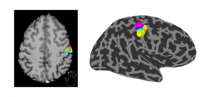
Our group has previously used UHF-fMRI to form spatial maps of how individual fingers are mapped within the primary somatosensory cortex of the human brain, Figure 1, and the finer somatosensory sub-areas previously only defined by histology. We now plan to publish a probabilistic atlas of the representation of the fingers in the primary somatosensory cortex and to show how these sensory maps are altered in the brains of patients with clinical conditions. We will also use ultra-high submillimetre spatial resolution fMRI to assess cortical microstructure and unravel the functional organisation of the cortex at the level of the six cortical layers to assess feed forward and feedback connectivity.
Another method the project is employing is magnetoencephalography (MEG). This is a non-invasive neuroimaging technique that measures magnetic fields at the scalp surface from which brain activity can be mapped. Using MEG, activity in local neuronal populations can be inferred and facilitate the assessment of long-range communication between cortical regions with unprecedented temporal resolution. The combination of UHF-fMRI and MEG data yields cortical mapping with uniquely high spatial and temporal precision allowing the assessment of how brain function is altered between healthy controls and patients.
For a further understanding of the neuroscience of touch, the project collaborates with Professor Johan Wessberg and colleagues from University of Gothenburg, Sweden on the use of intraneural microstimulation (INMS). In INMS, a single nerve fibre can be stimulated electrically using a very small current. This involves inserting an extremely fine needle through the skin and into an underlying nerve, allowing unprecedented access to the earliest stages of information transfer to the brain. This effect enables a person to feel touch without there being any actual skin stimulus. By combining INMS with neuroimaging techniques of UHF-fMRI and MEG, it is possible to resolve the significance and impact of single sensory nerve inputs and bridge an important scientific and clinical gap between input codes in mechanosensitive afferents and their dynamic interaction to generate a percept.
Neurological disorders are the most common cause of disability, with 10 million sufferers in the UK. Disturbances of somatic sensation are a major contributor to such disability. Specifically, in this project, we are studying the two clinical conditions of focal hand dystonia and carpal tunnel syndrome, which have been specially selected to provide new understanding of somatosensory neuropathology. We plan to use UHF-fMRI to assess how finger representations are altered in somatosensory and motor cortex regions and how the brain re-maps following therapeutic interventions. Sensory deficits are prominent in several conditions, including neurotraumatic injury, neurodegeneration and complex regional pain syndrome. The results expected might help to identify the underlying neuronal mechanisms and to develop novel treatments, improving the lives of many in the process.
Final remarks
Using UHF-fMRI and MEG we aim to advance understanding of human somatosensation and perception. We aim to address how the brain encodes and integrates touch and how the patient’s brain re-maps various motor and somatosensory pathways.
These projects are performed in collaboration with University of Gothenburg, Sweden (Lead: Professor Johan Wessberg) and Liverpool John Moores University (Lead: Professor Francis McGlone) and are funded by the Medical Research Council (Grant number MR/M022722/1).
Further reading
Sanchez-Panchuelo, R. M., Besle, J., Beckett, A., Bowtell, R., Schluppeck, D., and Francis, S. (2012). Within-Digit Functional Parcellation of Brodmann Areas of the Human Primary Somatosensory Cortex Using Functional Magnetic Resonance Imaging at 7 Tesla. J. Neurosci. 32, 15815–15822. doi:10.1523/JNEUROSCI.2501-12.2012.
Sanchez-Panchuelo, R. M., Francis, S., Bowtell, R., and Schluppeck, D. (2010). Mapping Human Somatosensory Cortex in Individual Subjects With 7T Functional MRI. J. Neurophysiol. 103, 2544–2556. doi:10.1152/jn.01017.2009.
Sanchez Panchuelo RM, Ackerley R, Glover PM, Bowtell RW, Wessberg J, Francis ST, McGlone F. Mapping quantal touch using 7 Tesla functional magnetic resonance imaging and single-unit intraneural microstimulation. Elife. 2016 May 7;5. pii: e12812. doi: 10.7554/eLife.12812.
Glover PM, Watkins RH, O’Neill GC, Ackerley R, Sanchez-Panchuelo R, McGlone F, Brookes MJ, Wessberg J, Francis ST.An intra-neural microstimulation system for ultra-high field magnetic resonance imaging and magnetoencephalography. J Neurosci Methods. 2017 Oct 1;290:69-78. doi: 10.1016/j.jneumeth.2017.07.016.Epub 2017 Jul 23.
Please note: this is a commercial profile
Professor Susan Francis
Professor
Sir Peter Mansfield Imaging Centre,
University of Nottingham
Tel: +44 115 846 6518
susan.francis@nottingham.ac.uk


