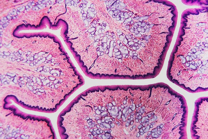
Recent years have seen an increase in techniques developed for spatial transcriptomics, enabling gene expression patterns to be uncovered within intact, three-dimensional tissues.
As these tools become more refined, efforts are beginning to integrate additional molecular markers analyses. This essay will list some of these examples and discuss the future of this research direction.
One day it will likely be possible to use a single analytical pipeline to extract proteomic, transcriptomic and genomic information from tissues in a high throughput and spatially defined manor. As these tools come into fruition, researchers will then be equipped to gain a better understanding of fundamental biology, as well as mechanisms of disease.
Thus far, sequencing-based in situ multiomics has not been demonstrated in tissue samples. Success, however, has been demonstrated in cell culture. In 2021, the group of Long Cai at Caltech demonstrated the readout of proteins, RNA and genomic DNA in cells using an in situ hybridisation approach (1).
For RNA and genomic readout, nucleic acid capture and readout was performed using DNA probes designed to bind to their respective molecular complement. For the detection of proteins, antibodies against targets were modified to contain a DNA strand unique to each target. This general strategy is an effective means to convert a protein detection mechanism into a nucleic acid one, thereby unifying the readout modality. It is unclear if this technique can be immediately adapted to tissue or if optimisation would be necessary. In general, the complexity of the three-dimensional environment, as well as the presence of extracellular matrix, make it more challenging to perform such analytical techniques.
What does the future hold for this area of research? It is expected that spatial sequencing approaches will also be used to demonstrated in situ multiomics (2). For instance, ExSeq enables super resolution imaging of transcriptomes by combining in situ sequencing with expansion microscopy (3). ExSeq has been used to in combination with antibodies, however not in a highly multiplexed way. This could easily be overcome by using antibodies labelled with unique oligonucleotides such as was used for the original expansion microscopy demonstration (4), as well as other examples (5). Integration of information about the genome could also be captured using variations of techniques that have already been developed (6,7).
Interestingly, the advent of synthetic biology techniques has opened up the possibility to encompass the analysis of modalities that are natively found in tissue but rather written there by researchers. Two areas where this will be possible are the study of development, as well as brain connectivity. For both areas, scientists have invented ways to write signatures into tissues that can be read out at a later time. For instance, transgenic mice have been engineered to use CRISPR to mutate and create a record of each cell’s development (8). For brain anatomy, viral vectors have been created that label cells with nucleic acid identifiers and inform about cell connectivity (9). By creating methods that can capture and read out these sequences, connectomics and cell lineage can be read out along with more classical OMICs information.
There is currently an explosion of effort focused on creating improved tools to study tissues. The field has made tremendous progress improving the ability to readout specific molecular types with high throughput. The future now lies on integrating these disparate protocols into a cohesive pipeline that can interrogate multiple modalities simultaneously. It is currently the dawn of in situ multiomics. The next years will be filled with innovation and future generations will have improved generations of technology that will unlock knowledge of biology that is not obtainable today.
References
(1) Takei, Y. et al. Integrated Spatial Genomics Reveals Global Architecture of Single Nuclei. Nature 2021, 590, 344-350.
(2) Turczyk, B. M., Busby, M., Martin, A. L., Daugharthy, E. R., Myung, D., Terry, R. C., Inverso, S. A., Kohman, R. E., Church, G. M. Spatial Sequencing: A Perspective. J Biomol Tech 2020, 31 (2), 44-46.
(3) Alon, S. et al. Expansion Sequencing: Spatially Precise in situ Transcriptomics in Intact Biological Systems. Science 2021, 371, (6528), eaax2656.
(4) Chen, F., Tillberg, P. W., Boyden, E. S. Expansion Microscopy. Science 2015, 347, 543-548.
(5) Kohman, R. E. and Church, G.M. Fluorescent in situ Sequencing of DNA Barcoded Antibodies. bioRxiv 2020, doi: https://doi.org/10.1101/2020.04.27.060624.
(6) Nguyen, H.Q. et al. 3D mapping and Accelerated Super-resolution Imaging of the Human Genome using in situ Sequencing. Nat. Methods, 2020, 17, 822–832.
(7) Payne A.C. et al. In situ Genome Sequencing Resolves DNA Sequence and Structure in Intact Biological Samples. Science 2020, 371, (6532), eaay3446.
(8) Kalhor, R., Kalhor, K., Mejia, L., Leeper, K., Graveline, A., Mali, P., Church, G.M. Developmental Barcoding of Whole Mouse via Homing CRISPR. Science 2018, 361, (2018), eaat9804
(9) Zador, A.M. Dubnau, J., Oyibo, H.K., Zhan, H., Cao, G., Peikon, I.D. Sequencing the Connectome. PLOS Biology 2012, 10(10): e1001411.
Please note: This is a commercial profile
© 2019. This work is licensed under CC-BY-NC-ND.


