Anna Knuuttila, Senior Scientist and Kaisa Helminen, CEO of Fimmic explain how artificial intelligence (AI) empowers pathologists when it comes to cancer care
By 2030, nearly 22 million new cancer cases are expected and the burden on the healthcare system to quickly and accurately analyse the tissue samples to diagnose them and develop lifesaving drugs is immense. To help address the growing number of samples from cancer, as well as other diseases, Fimmic, a Finnish software company, has developed Aiforia™ Cloud. It combines deep learning artificial intelligence (AI) image analysis cloud solution and automatised pathology image analysis capabilities. With this Fimmic aims to improve the speed and accuracy of tissue-based diagnostics and thus enable efficient, precise and more personalised care.
Formerly known as WebMicroscope, Aiforia Cloud is already used by more than 6,000 pathologists, researchers and pharmaceutical R&D teams to manage and share digital slide collections. With the new Aiforia platform CNN (convolutional neural network) deep learning algorithms can be easily developed in just days or less, compared to the months needed for conventional machine vision approaches. Along with the algorithms being easy to create, the entire platform itself is simple, easily accessible and affordable with SaaS model. Users can deploy and use the platform through a browser.
For the first time, the Aiforia deep learning algorithm can mimic the human observer in understanding the context in tissue and it can automatically and accurately perform laborious image analysis tasks in a fraction of the time. This gives users significant time back in their day for high-value work, such as analysing more complex or rare samples.
“Digitising the field of pathology has been an important step forward for healthcare, but we’re once again poised for real disruption in the space,” said Kaisa Helminen, Fimmic CEO. “An ageing population and rise in cancer prevalence, coupled with the radical innovation of AI technology, have created both a significant need and opportunity for digital pathology to evolve. At Fimmic, we are driven by the common goal of better and more accurate medical analysis, diagnoses and treatments and we are proud to offer the first solution of its kind to bring deep learning AI to the fingertips of all pathologists and researchers, empowering users to explore and discover more with less time, money and effort.”
Application example: Quantification of Ki67 in breast cancer
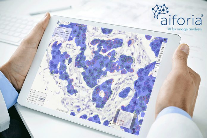
Fimmic has developed deep learning algorithms for several different applications, for example, in drug development and in research on cancer, neurodegenerative diseases and liver diseases, but this technology can be applied to numerous other applications as well. One example of the developed algorithms is epithelium segmentation and quantification of Ki67 positive and negative cells in breast tumour sections. Ki67 is a nuclear protein that is expressed in proliferating cells and may be used for the estimation of prognosis of breast cancer, prediction of responsiveness to therapy, estimation of residual risk and treatment efficacy1.
Unbiased and reproducible quantification of Ki67 positive and negative tumour cells is important both for breast cancer research, as well as in clinical practice. The Ki67 scoring methods are traditionally based on visual assessment1. This is laborious and time-consuming and subject to human error. Since it is not possible to count all Ki67 positive and negative cells from the whole tumour area, the counting by eye is always an approximation. In the novel deep learning algorithm these problems have been overcome.
Furthermore, unlike algorithms that have been generated with conventional machine vision, the deep learning algorithms can automatically segment the tumour epithelium from the stroma and count the Ki67 positive and negative cells in the right context, resulting in significantly faster and more reproducible results, with no hands-on-time. The Ki67 counting is performed from the digitised breast tissue sections. There is no need to acquire any local hardware for image processing, since the Aiforia deep learning algorithms are available as a service from a cloud platform, accessible with any modern browser. See our video.
The benefits are:
- Fully automated analysis from the whole tumour area;
- Analysis from the area of interest. No double-staining needed for epithelium/stroma segmentation;
- Fast, accurate and reproducible results and;
- No local hardware or software needed.
About Fimmic
Fimmic, founded in 2013, is a spin-off company from the Finnish Institute for Molecular Medicine at the University of Helsinki. Fimmic’s unique Aiforia Cloud brings together deep learning-based image analysis and high-performance cloud computing. The SaaS solution enables fast, accurate and affordable analysis support for every pathologist and medical researcher in tissue-based image analytics through a zero friction cloud deployment and intuitive online user interface.
New algorithms can be ordered on-demand or with a self-service model where users can generate their own deep learning algorithms by training convolutional neural networks to learn, detect and quantify specific features of interest in tissue images. Aiforia is currently sold for research use. Please visit www.aiforia.com for more information.
References
1 Dowsett et al. Assessment of Ki67 in Breast Cancer: Recommendations from the International Ki67 in Breast Cancer Working Group. JNCI 2011;103: 1656–1664, https://doi.org/10.1093/jnci/djr393
Please note: this is a commercial profile
Anna Knuuttila
Senior Scientist, Fimmic
Kaisa Helminen
CEO, Fimmic
Tukholmankatu 8contrib
00290 Helsinki
Finland


