Open Access Government produces compelling and informative news, publications, eBooks, and academic research articles for the public and private sector looking at health, diseases & conditions, workplace, research & innovation, digital transformation, government policy, environment, agriculture, energy, transport and more.
Home 2026
Archives
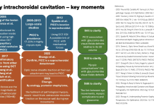
Peripapillary intrachoroidal cavitation and biomechanical considerations: A multi-stage narrative
Dr Adèle Ehongo discusses Peripapillary intrachoroidal cavitation and biomechanical considerations in a multi-stage narrative.
OCT: A practical tool for diagnosing buried optic disc drusen
Dr Adèle Ehongo addresses the diagnosis of buried optic disc drusen (BODD) using Optical Coherence Tomography (OCT) and the importance of correlating visual field abnormalities and OCT data, especially in normal tension glaucoma, to distinguish it from Optic Disc Drusen (ODD).
Peripapillary intrachoroidal cavitation and myopic peripapillary changes
Peripapillary intrachoroidal cavitation and myopic peripapillary changes: Optical Coherence Tomography analysis.
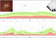
Peripapillary Intrachoroidal Cavitation, a masquerade of normal-tension glaucoma
Dr Adèle Ehongo discusses peripapillary intrachoroidal cavitation (PICC), a masquerade of normal-tension glaucoma.
Understanding the link between PICC and myopic complications
Dr Adèle Ehongo discusses the pathogenesis of peripapillary intra-choroidal cavitation and its implications for myopic complications.
Glaucoma clinic within the ophthalmology department
Professor Adèle Ehongo discusses her work in context of the ophthalmology department at Brussels University Hospital.
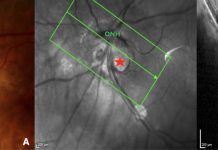
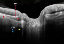
Spotting peripapillary intra-choroidal cavitation using OCT
Adèle Ehongo explores the potential of optical coherence tomography for diagnosing peripapillary intra-choroidal cavitation in myopic eyes.

