Home 2024

Archives

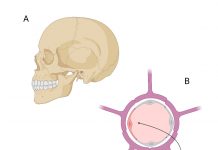
Figure 1: The central nervous system (CNS) is protected from mechanical injury by the skull (A) and from chemical noxious agents by the blood-brain barrier (B, C). The latter consists of the endothelial barrier in most parts of the CNS (B) and the tanycytic barrier in the hypothalamus (C). We develop gene vectors transducing endothelial cells and tanycytes to treat genetic and non-genetic diseases of the brain.

Bridging brain barriers for gene therapy

Reflecting on the challenges in treating brain diseases, this article explores ways to transduce the blood-brain barrier as well as the critical role of tanycytes as a target for gene therapy vectors.
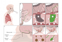
Figure 1. Two routes of brain infection by which SARS-CoV-2 may enter the hypothalamus and infect GnRH neurons. The virus attaches to and enters cells harbouring surface receptors such as ACE2 (black spanner-like symbol) or NRP1 (red cylinder). GnRH neurons exhibit both, which might increase their vulnerability. A. The haematological route, whereby the respiratory virus (white spheres) makes its way through the lungs into the bloodstream (dark red), and thence into the median eminence (ME), a part of the hypothalamus that harbours ”fenestrated” or leaky blood vessels. The virus may affect a number of different cell types locally, including GnRH neurons (green), whose secretory terminals approach the fenestrated vessels, and tanycytes (grey), whose processes control this secretion and also transport other bloodborne hormones and other substances into the brain. Once inside the brain ventricles (V), fluid-filled canals within the brain, the virus can also travel to other areas. B. The olfactory route, whereby the virus enters the olfactory bulb (OB) of the brain directly from the nose across the bony cribriform plate by means of nerve bundles or infected olfactory neurons and other cell types. GnRH neurons, which are born in the nasal epithelium during the embryonic period and migrate into the brain along these nerve bundles to their final positions, still maintain a connection with their birthplace, and could be thus be infected directly or indirectly through olfactory neurons or other cells. GnRH neurons also project to parts of the brain involved in higher functions such as cognition, potentially contributing to long-COVID symptoms such as “brain fog”.

Brain infection by SARS-CoV-2: Lifelong consequences

The WATCH team, founded to elucidate the role played by specialized brain cells called tanycytes in various physiological processes, has been investigating how and where the SARS-CoV-2 virus infects the brain, and some long-term consequences of this neuro-invasion.
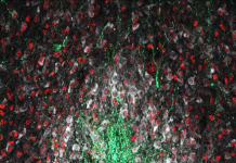
Image showing NO-producing neurons (white) interacting with other key neuronal populations shaping minipuberty in the hypothalamus (green: gonadotropin-releasing hormone neurons (GnRH), red: estrogen receptor-alpha expressing neurons (Erα)) https://doi.org/10.1016/j.freeradbiomed.202 2.11.040, minipuberty

The miniNO project: Helping to minimize risks of premature births

Associative mechanisms linking a defective minipuberty to the appearance of mental and non-mental disorders: infantile NO replenishment as a new therapeutic possibility.
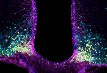
image showing tanycytes in purple and the neurons they interact with in the hypothalamus (yellow: appetite promoting neurons expressing neuropeptide Y (NPY); blue: appetite suppressing neurons expressing the propopiomelanocortin (POMC))

The WATCH project: Tanycytes in health and disease

The WATCH project aims to elucidate how tanycytes mediate physiological processes by acting as gatekeepers between the brain and body, how their dysfunction is involved in various disorders and age-related impairments, and what can be done to prevent or correct these.

Follow Us

Advertisements