Elize Sturkenboom – van de Wetering, Venture Leader of Lung Oncology at Philips Healthcare, discusses the diagnosis and treatment of lung cancer through new minimally invasive therapy
Lung cancer kills around 1.8 million people in a year worldwide. It is the leading cause of cancer death globally, accounting for greater loss of life than breast, colon, and prostate cancer combined (1). While early diagnosis and treatment are critical to better outcomes and quality of life, the majority of lung cancers are currently diagnosed too late, with minimal chance of a surgical cure. Only 21% of all people diagnosed with lung cancer will survive 5 years or more. When lung cancer patients are diagnosed early and treated immediately, a patient’s chance of survival at 10 years jumps to 92%.(2)
New hope on the horizon for lung cancer screening
Changes in healthcare policies and government involvement have increased lung cancer screening. The implementation of lung cancer screening programs with low-dose CT scans are driving a shift resulting in more patients with curable lesions.
This comes with its own challenges. Small peripheral nodules are typically more complex to reach and localise during biopsy and surgery. The good news is that more and more physicians are adopting new techniques to diagnose and later treat these tiny lesions that are so difficult to catch.
The impact of new image guidance technology on early diagnosis and treatment
More and more accurate and less invasive methods are being introduced and adopted in clinical practice and have started a new era. These days, even barely visible nodules of just 8mm can be diagnosed and treated through new minimally invasive techniques. Advanced imaging integrated into operating rooms and bronchoscopy suites can be combined with navigation bronchoscopy to achieve high precision diagnosis and fulfil the need to combine diagnosis and staging with minimally invasive therapy in one room.
“Cone-beam CT is the critical step towards targeting sub-20 mm nodules and an essential tool for the transition towards bronchoscopic microwave ablation of peripheral lung lesions,” said Professor Shah Pallav, MD., consultant respiratory physician at Royal Brompton Hospital in London, UK.
Diagnosis of small peripheral lung lesions
Recently, Radboud university medical center (Radboudumc), a leading Dutch academic research and teaching hospital (Nijmegen, the Netherlands), have announced the positive results of a clinical study aimed at setting a new standard of safety and accuracy in the diagnosis of small peripheral lung lesions. The study has set a new milestone with 90% diagnostic accuracy of lung nodules suspicious of cancer using Philips Lung Suite technology combined with the company’s Image Guided Therapy System – Azurion for bronchoscopy procedures.
Philips’ solution uses real-time 3D imaging with augmented fluoroscopy to support high precision diagnosis and minimally-invasive therapy in one room. The results of the study were published in the October 2021 issue of the Journal of Bronchology & Interventional Pulmonology: Volume 28 – Issue 4.
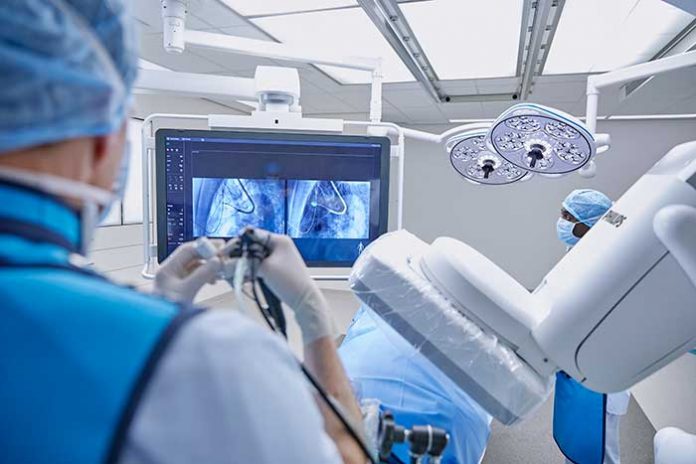
Bronchoscopy procedure performed with Philips advanced imaging
This Philips’ platform also offers the promising outlook to not only diagnose but immediately treat these lung cancer patients using novel procedures such as tumor ablation.
Dr. Kelvin Lau, consultant and lead thoracic surgeon at St Bartholomew’s Hospital, London, successfully diagnosed and treated lung cancer patients using cone beam CT for biopsy, bronchoscopic microwave ablation, all in one procedure, during initial clinical trial.
Advanced imaging with Philips Lung Suite is used during procedures for real-time 3D image guidance and confirmation. Additional clinical trials at various hospitals are expected to start soon.
All-in-one lung cancer diagnosis and treatment
Philips Lung Suite enables all-in-one lung cancer diagnosis and treatment. It provides advanced real-time 3D imaging with augmented fluoroscopy on the company’s Image Guided Therapy System – Azurion, combined with dedicated software. With Philips’ Cone Beam CT imaging, the X-ray detector rotates around the patient to generate a CT-like image in around 5 seconds, providing clinicians with a high-resolution 3D view of the target lesion and other anatomical structures. This allows the clinician performing the biopsy procedure to be continually guided by high-quality real-time imaging to advance a catheter towards the lesion through a bronchoscope. Once done, its position can be confirmed in real-time using the same imaging modality and a biopsy sample removed.
Philips has a comprehensive portfolio of lung cancer diagnosis and treatment solutions. In addition to Philips Lung Suite, the company’s Lung Cancer Orchestrator provides an integrated lung cancer patient management system for both CT lung screening programs and incidental pulmonary findings programs that manages and monitors patients every step of the way in their lung cancer screening and treatment decision journey.
Several centers around the globe have implemented this cutting-edge technology and innovating procedure with the aim to shorten the patient pathway, improve first time right diagnosis and reduce the overall cost of care.
“Philips aspires to transform lung cancer from a deadly disease to a managed condition, by leaving no patient behind with early-stage lung cancer. Philips was there when cardiac surgery transformed into coronary stenting, and are here right now to drive that change for lung cancer patients,” tells Elize Sturkenboom, Venture Leader IGT Lung Oncology.
The knowledge and the technology are now available to diagnose and treat lung cancer earlier, more accurately and less invasively than ever. It’s now time to move the needle and make a difference.
References
[1] https://www.who.int/news-room/fact-sheets/detail/cancer
[2] https://www.cancer.org/cancer/lung-cancer/detection-diagnosis-staging/survival-rates.html
Please note: This is a commercial profile
© 2019. This work is licensed under CC-BY-NC-ND.


