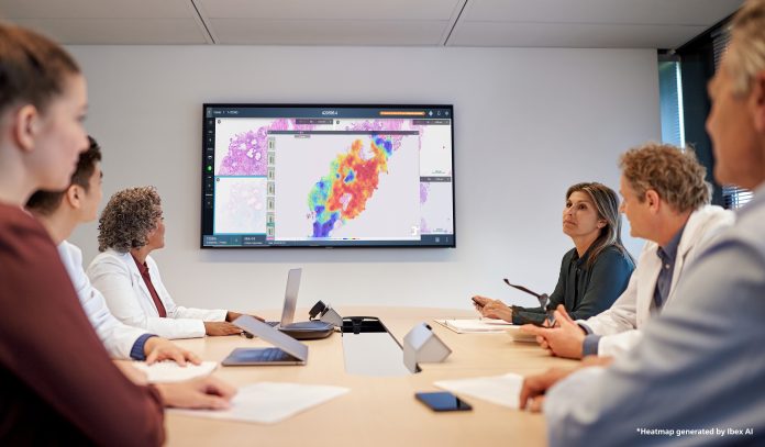
Philips explains how digital pathology accelerates the pathway to personalized cancer care
Pathologists play a crucial role in cancer diagnosis and treatment. Given that it is projected that more than 35 million new cancer cases will be diagnosed in the year 2050 – a 77% increase from the estimated 20 million cases in 2022 (1) – the demand for pathology services will continue to rise. However, the number of pathologists is critically low. It is estimated that globally, four times more pathologists are needed than are currently in practice. (2)
Unlike many hospital departments that have migrated to digital workflows, most pathology departments use an analog process, viewing tissue samples on glass slides that must be couriered to multidisciplinary team members or for second opinions. This creates inefficient workflows and barriers to data access. The adoption of digital pathology for primary diagnosis is estimated to be less than 10% in the US (3) and 23% in Europe and Asia. (4)
Digital pathology offers efficiency and enhanced collaboration
Growing caseloads, fewer pathologists, workflow challenges, and the need for timely, precise diagnosis are driving the adoption of digital pathology. Digital pathology redefines diagnostic workflows with high-resolution imaging, the ability to integrate artificial intelligence (AI) and remote reading and consultation. With digital pathology, pathologists can access and share information more efficiently, route cases to other members of the multidisciplinary care team, obtain second opinions more quickly, and improve overall diagnostic turnaround times, ultimately leading to more personalized cancer care.
High-volume digital pathology laboratories can offer telepathology services, which benefit hospitals without on-site pathologists or who are working beyond capacity by allowing them to offload cases without the time, expense, and delay of manually transporting slides.
The analog to digital transformation
Philips introduced the first digital pathology solution cleared by the FDA for clinical use in 2017. Since then, Philips has helped more than 300 customers worldwide implement digital pathology and over 38 million slides have been digitized on these systems. These implementations and deep experience have uncovered best practices for a smooth transition from analog to digital pathology.
First, a digital pathology solution should include user-centric intuitive interfaces that make the transition from microscope-based workflows easy. Second, effective educational programs, including training on slide digitization, help pathology labs adopt the new technologies. Comprehensive training sessions, often using the lab’s own slides, validate the solution and ensure that pathologists feel comfortable and confident in their new digital environment. Philips supports these training sessions with change management, workflow analysis, and optimization to help pathology departments successfully transform quickly and with as little stress as possible.
An ideal digital pathology system will also integrate effectively with existing Laboratory Information Systems to ensure relevant data is consistent throughout the care pathway. It will also enable AI applications* that ease pathologists’ workload by prioritizing cases, highlighting areas of concern, and facilitating reporting.
Once digital pathology systems are in place, it is essential to leverage the insights gained from their use and identify opportunities for further workflow enhancements. This ongoing evaluation ensures that the digital systems are utilized to their fullest potential, unlocking efficiency and diagnostic accuracy.
Productivity gains and the power of AI
Feedback from system users is highly positive; 100% of pathologists surveyed said going digital helps reach diagnostic consensus (5) and studies show time savings of up to 19 hours per day (5) on case logistics and reading.
With one of the largest installed bases in the world, Philips has helped pathology labs reach impressive results by transforming to digital pathology workflows. Staff adapt to the new digital system within eight days after only six hours of training and realize productivity gains of 25%. (5) When AI is added, it can boost productivity by up to 37%. (6) In cases of prostate cancer, AI reduces the turnaround time to diagnosis from 1.8 days to 9.4 minutes (6) compared to the standard of care using a microscope. AI also has shown high accuracy in identifying pathology across tissue types. (7,8)
Our digital pathology systems have years of performance in clinical use and have enabled pathology labs to institute new, more efficient ways of working. For example, Ohio State University, a pioneer in digital pathology, uses the Philips digital pathology solution for clinical care, research, and telepathology, averaging more than 2,300 scans per day. (9) Paris Saint- Joseph & Marie Lannelongue Hospitals, use its Philips digital pathology solution to support multidisciplinary tumor boards in guiding personalized care pathway selection, and there are many more use cases around the globe. (9)
Transforming care
Transformation to a fully digital pathology workflow improves laboratory operations and enhances a pathologist’s efficiency. It enables streamlined image analysis, remote collaboration, and faster diagnosis, opening new opportunities for research and clinical collaboration. Adopting digital pathology is vital for pathology laboratories in supporting clinical teams that develop the best possible path for every patient, ensuring personalized, accessible cancer care.
*PIPS enables iSyntax files, and with the Software Development Kit (SDK), third-party companies can use this for AI capabilities
References
- Global cancer burden growing, amidst mounting need for services. World Health Organization news release. 1 February 2024.
- Bychkov, A., and Fukuoka, J. Evaluation of the global supply of pathologists. United States & Canadian Academy of Pathology 111th Annual Meeting. March 2022.
- Lloyd M. Going digital, turning green. The Pathologist. 25 September 2024.
- Pinto DG, Bychkov A, Tsuyama N, Fukuoka J and Eloy C. Real-world implementation of digital pathology: results from an intercontinental survey. Laboratory Investigation. December 2023; 103(12).
- Survey of 52 pathologists, lab managers, and lab technicians in Europe, 2018.
- Information provided by Ibex AI. Raoux et al. Modern Pathology (2021) 34 (suppl 2): 598-599.
- Information provided by Ibex AI. Sandbank et al., npj Breast Cancer, December 2022
- Information provided by Ibex AI. Sandbank et al., Modern Pathology 2022, 35,513-514
- Results are specific to the institution where they were obtained and may not reflect the results achievable at other institutions.
The Ibex platform is for Research Use Only (RUO) in the United States and pending 510K.

This work is licensed under Creative Commons Attribution-NonCommercial-NoDerivatives 4.0 International.


