The role of ultrasound for Inferior Vena Cava measurement (IVC) in patients presenting with shortness of breath is often debated. Authors have disputed different modes and points of measurement and with varying probe placement.1-5 Additionally, patient position, habitus, degree of respiratory distress, and the presence of mechanical ventilation can influence the size and collapsibility of the IVC. Common agreement may be found from a recent metanalysis suggesting a moderate level of evidence supporting the IVC diameter is low in hypovolemic patients as compared with euvolemic patients.6
The caval index calculates the percentage collapse of the IVC: IVC expiratory diameter – IVC inspiratory diameter, divided by the IVC expiratory diameter x 100 = caval index (%). In the setting of shortness of breath, a caval index near 100% suggests complete collapse of the IVC and is indicative of volume depletion. The closer the number to 0% the more likely the patient has intravascular volume overload.7 Additionally, cardiac tamponade from pericardial effusion should be considered with a non-collapsable IVC in patients who present with shortness of breath. The BRIPPED scan is a screening tool for patients with shortness of breath of unclear etiology. Among its components discussed below, the scan simplifies the caval index by qualitatively evaluating the collapse of the IVC.
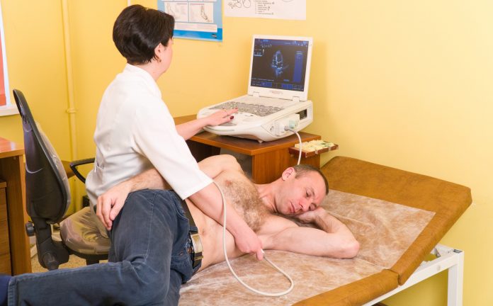
The IVC is visualised in the long axis plane in patients who are semi-recumbent or supine. The IVC should be visualised as it enters the right atrium, to differentiate it from the aorta that runs parallel to the IVC. With the BRIPPED protocol, the sonographer may image the IVC, and obtain cardiac windows using the same lower frequency phased array probe to evaluate ejection fraction and pericardial effusion. The probe is placed below the xiphoid bone, and the probe marker rotated towards the patient’s head. Alternatively, the probe may be placed anterior to the mid axillary line, with the probe marker towards the head.
BRIPPED Protocol:
The BRIPPED scan is an effective screening tool for shortness of breath that evaluates pulmonary B-lines, Right ventricle size and strain, Inferior Vena Cava (IVC) collapsibility, Pleural and Pericardial Effusion, Pneumothorax, Ejection Fraction of the left ventricle, and lower extremity Deep Venous Thrombosis.
B-lines: Sonographic pulmonary Blines have been shown to correlate with congestive heart failure.8-11, 15, 16 A high frequency linear probe is used to evaluate at minimum 2 mid clavicular apical lung windows.
RV strain: Right ventricular (RV) enlargement can be caused by a Pulmonary Embolus (PE), acute RV infarct, Congestive Heart Failure (CHF), pulmonary valve stenosis or pulmonary hypertension, and is a risk factor for early mortality in PE.17 A low frequency phased array probe is used to evaluate RV strain in an apical 4 chamber view.
IVC-size and collapsibility: Using an IVC size cut off of 2.0 cm has been shown to have a sensitivity of 73% and specificity of 85% for a Right Atrial Pressure (RAP) above or below 10 mmHg. The collapsibility during forced inspiration of less than 40% has even greater accuracy for elevated RAP (sensitivity 91%, specificity 94%, NPV 97%).18 A low frequency phased array or curvilinear probe is used to visualise the IVC long axis, and dynamic imaging is used to assess collapsibility as either complete or less than 40%.
Pneumothorax: Bedside ultrasound is more accurate than supine chest xray with diagnostic ability approaching that of CT. 19, 20 The same windows for B-lines are utilised for pneumothorax screening. Additionally any area of decreased breath sounds, or crepitus palpated along the chest wall is evaluated for pneumothorax with a high frequency linear probe.
Pleural effusion: EUS has been shown to have an accuracy similar to a CXR for evaluation of pleural effusion.13, 14 A low frequency phased array or curvilinear probe is used to evaluate each mid axillary line at the costophrenic angle in the sitting patient.
Pericardial effusion: EUS has a sensitivity of 96% and specificity of 98% compared to formal echocardiography. 21 A low frequency phased array probe is used to evaluate pericardial effusion from an apical 4 chamber view and a parasternal long axis view of the heart.
EF: The qualitative assessment of left ventricular ejection fraction by emergency physicians has been shown to correlate well with an assessment by a cardiologist.22-24 The same low frequency probe and parasternal long axis used to evaluate pericardial effusion is used to evaluate ejection fraction. Dynamic qualitative assessment of ejection fraction is classified as normal, depressed, or severely depressed.
DVT in lower extremities: Ultrasound was performed by emergency physicians using a two point compression venous ultrasound on patients with suspected lower extremity DVT. This approach had a 100% sensitivity and 99% specificity in diagnosing DVT, compared to a reference venous ultrasound in radiology.25 A high frequency linear probe evaluates compressibility of the common femoral and popliteal fraction. Dynamic qualitative assessment of ejection fraction is classified as normal, depressed, or severely depressed.
DVT in lower extremities: Ultrasound was performed by emergency physicians using a two point compression venous ultrasound on patients with suspected lower extremity DVT. This approach had a 100% sensitivity and 99% specificity in diagnosing DVT, compared to a reference venous ultrasound in radiology.25 A high frequency linear probe evaluates compressibility of the common femoral and popliteal probe to evaluate the parasternal long axis and apical 4 chamber, noting the presence or absence of pericardial effusion, ejection fraction, and RV strain. Then the long axis of the IVC is evaluated for dynamic collapsibility. Moving laterally, the costophrenic angles are evaluated bilaterally for pleural effusion. The probe is switched to the high frequency probe to evaluate each lung apex is evaluated in the mid clavicular line for the presence of pneumothorax and B lines. Lastly, the dynamic 2 point DVT screening is performed with compression ultrasound. The BRIPPED protocol and other bedside ultrasound resources can be viewed here:
References:
1 Kircher B, Himelman R, Schiller N. Noninvasive estimation of right atrial pressure from the inspiratory collapse of the inferior vena cava. AM J Cardiol 1990; 66: 493-6.
2 Akilli B, Bayir A et al. Inferior vena cava diameter as a marker of early hemorrhagic shock: a comparative study. Ulus Travma Acil Cerrahi Derg 2010;16(2):113-8.
3 Barbier C, Loubières Y, Schmit C, Hayon J, Ricôme JL, Jardin F, Vieillard-Baron A. Respiratory changes in inferior vena cava diameter are helpful in predicting fluid respon- siveness in ventilated septic patients. Intensive Care Med 2004; 30:1740–1746
4 Blehar DJ, Dickman E, Gaspari R. Identification of congestive heart failure via respiratory variation of inferior vena cava. Am J Em Med 2009;27:71–5.
5 Blehar et al. Inferior vena cava displacement during respirophasic ultrasound imaging. Critical Ultrasound Journal 2012, 4:18
6 Dipti A et al. Role of inferior vena cava diameter in assessment of volume status: a meta-analysis. A JEM 2012 (30). 1414 -19.
7 Nagdev AD, Merchant RC, Tirado-Gonzalez A, et al. Emergency department bedside ultrasonographic measurement of the caval index for noninvasive determination of low central venous pressure. Ann. Emerg. Med. 2010;55:290-5.
8 Lichtenstein D, Meziere G, Biderman P, Gepner A, Barre O. The comet-tail artifact. An ultrasound sign of alveolar-interstitial syndrome. Am J Respir Crit Care Med. 1997; 156:1640–6.
9 Soldati G, Copetti R, Sher S. Sonographic Interstitial Syndrome The Sound of Lung Water. J Ultrasound Med 2009; 28:163-174.
10 Reibig A, Kroegel C. Trasnthoracic sonography of diffuse parenchymal lung disease: the role of comet tail artifacts. J Ultrasound Med. 2003;22:173-180.
11 Rumack CM, Wilson SR, Charboneau JW. Diagnostic Ultrasound. 3rd ed. St. Louis, MO: Mosby; 2004.
12 Copetti R, Cattarossi L, Macagno F, Violino M, Furlan R. Lung Ultrasound in respiratory distress syndrome: a useful tool for early diagnosis. Neonatology. 2008:94(1):52-9.
13 Wernecke K. Sonographic features of pleural disease. A JR AM J Roentgenol. 1997;168:1061-1066. 14 Vignon P, Chastagner C, Berkane V, et al. Quantitative assessment of pleural effusion in critically ill patients by means of ultrasonography. Crit Care Med. 2005;33:1757-1763.
15 Lichtenstein D, Meziere G. Relevance of lung ultrasound in the diagnosis of acute respiratory failure: the BLUE protocol. Chest. 2008;134:117-125.
16 Liteplo, A.S., et al., Emergency thoracic ultrasound in the differentiation of the etiology of shortness of breath (ETUDES): sonographic B-lines and N-terminal pro-brain-type natriuretic peptide in diagnosing congestive heart failure. Acad Emerg Med, 2009; 16(3):201-10.
17 Kucher, N., et al., Prognostic role of echocardiography among patients with acute pulmonary embolism and a systolic arterial pressure of 90 mm Hg or higher. Arch Intern Med, 2005; 165(15):1777-81.
18 Brennan, J.M., et al., Reappraisal of the use of inferior vena cava for estimating right atrial pressure. J Am Soc Echocardiogr, 2007; 20(7):857-61.
19 Kirkpatrick, A.W., et al., Hand-held thoracic sonography for detecting post-traumatic pneumothoraces: the Extended Focused Assessment with Sonography for Trauma (EFAST). J Trauma, 2004; 57(2): 288-95.
20 Xirouchaki N, Magkanas E, Vaporiid K, et al., Lung ultrasound in critically ill patients: Comparison with bedside chest radiography. Intensive Care Med, 2011; 37(9):1488-1493.
21 Mandavia, D.P., et al., Bedside echocardiography by emergency physicians. Ann Emerg Med, 2001; 38(4):377-82.
22 Alexander, J.H., et al., Feasibility of point-of-care echocardiography by internal medicine house staff. Am Heart J, 2004; 147(3): 476-81.
23 Moore, C.L., et al., Determination of left ventricular function by emergency physician echocardiography of hypotensive patients. Acad Emerg Med, 2002; 9(3):186-93.
24 Randazzo, M.R., et al., Accuracy of emergency physician assessment of left ventricular ejection fraction and central venous pressure using echocardiography. Acad Emerg Med, 2003; 10(9): 973-7.
25 Crisp, J.G., L.M. Lovato, and T.B. Jang, Compression ultrasonography of the lower extremity with portable vascular ultrasonography can accurately detect deep venous thrombosis in the emergency department. Ann Emerg Med, 2010; 56(6): 601-10.
Virginia M Stewart
MD RDMS RDCS RDMSK
Emergency Ultrasound Director, Emergency Ultrasound Fellowship Director
Department of Emergency Medicine
Tel: (757) 594 2000


